bloc 33rd Square Business Tools - viruses 33rd Square Business Tools: viruses - All Post
Showing posts with label viruses. Show all posts

Friday, April 22, 2016

Science to the Rescue! How Scientists Combat Viruses and Bacteria


Medicine  

New technology can provide opportunities to investigate the human biological processes at the cellular level, allowing them to discover innovative methods to prevent and fight infection by bacteria and viruses to create a healthier and longer-lived population.


Science is being used to solve one of the most ancient and persistent of problems, that of bacteria and viruses that cause disease, disability and death. New techniques are allowing researchers to see these life forms in minute detail and manipulate the genetic forces that cause them to proliferate. Here are just a few ways science is helping to conquer the problem of infection by these tiny predators.


Science Helping the Human Body Fight Bacteria

In one line of research, scientists are trying to harness the natural power of the human body to fight disease to create defenses against bacteria. They found that the body responds to the presence of a protein found in the tails, called flagella, of bacteria. Injecting the body artificially with this protein, flagellin, can trigger its natural defenses to fight diseases such as rotavirus, a common intestinal infection in children.

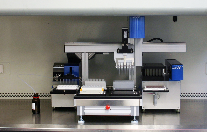
Utilizing the natural defenses of the body may help to reduce the use of antibiotics in the future, which cause the problem of antibiotic resistance. Researchers are aided in this technique by new types of computerized equipment, such as the Hudson compact SOLO-based ELISA workcell station, which can perform a series of reagent tests quickly and easily. These tests form the core of testing used for determining the ability of human tissues to produce antibodies that fight disease.
Related articles


Using Viruses to Fight Bacteria

Some scientists are learning to use viruses to fight bacterial infection. Viruses contain genomes called “bacteriophages,” which can be mixed and matched to target specific types of bacteria. When bacteriophages are introduced into the body, they can disrupt microbial communities, providing help for individuals with Crohn’s disease or other conditions caused by the presence of specific bacterial strains.
Hudson Robotics

DNA Gene Sequencing Tracks Virus Mutations

Unlike bacteria, which can reproduce easily, viruses must depend on cell mechanisms to help them copy themselves. Viruses have proteins on their surfaces that function as keys to attach to cell receptors, which allows them access to the internal mechanism of the cell. When the virus copies itself, changes often occur in the genetic material.

These changes are the mutations that allow genes to change constantly. DNA sequencing allows scientists to track these changes in the virus code, so that health professionals can determine the source of virus outbreak and how to stop it. This technology was used in 2013 to stop the spread of a hepatitis-A outbreak that was sourced to food contamination.

New technologies are sure to present even more opportunities to investigate the human biological processes at the cellular level, allowing them to discover innovative methods to prevent and fight infection by bacteria and viruses to create a healthier and longer-lived population.


By Anica OaksEmbed


Author Bio - A recent college graduate from University of San Francisco, Anica loves dogs, the ocean, and anything outdoor-related. She was raised in a big family, so she's used to putting things to a vote. Also, cartwheels are her specialty. You can connect with Anica here.


Friday, October 9, 2015

New Medical Test Can Diagnose Any Viral Infection


Medicine  


Before physicians can treat an ailment, they need to properly diagnose it. This can be particularly tricky with some illnesses, considering there are a lot of overlapping symptoms at times. A new medical tool called the ViroCap may change all of that.


Before physicians can treat an ailment, they need to properly diagnose it. This can be particularly tricky with some illnesses, considering there are a lot of overlapping symptoms at times. However, a new medical tool called the ViroCap may change all of that.

The ViroCap test can identify just about any type of viral infection patients may be suffering from, allowing doctors to correctly diagnose patients even when they don’t have any clues or ideas as to what to search for.

The Lowdown on the ViroCap

A study published in the Genome Research journal discusses the ViroCap test in detail. It can detect any virus with the capability of infecting humans or animals, from common to rare. That includes the full spectrum of deadly outbreaks, including Marburg virus, Ebola, severe acute respiratory syndrome (SARS) and more common outbreaks such as rotavirus and even norovirus.

What’s more interesting is the test can detect individual strains of viruses. Taking that one step further, it can even detect when someone is suffering from multiple viruses simultaneously. The latter point is important because there is currently no other test that can do such a thing.

But just how accurate is the ViroCap test? Currently, viruses are diagnosed using the polymerase chain reaction (PCR) test. Apparently, the ViroCap test has been reported to be 52 percent more accurate than the PCR testing method. That’s a big deal since the PCR test is the current industry “gold standard.”


Accurate but Unavailable

The author of the study, Todd Wylie, confirms the ViroCap test is remarkably accurate. He says, "The test is so sensitive that it also detects variant strains of viruses that are closely related genetically. Slight genetic variations among viruses often can't be distinguished by currently available tests and complicate physicians' ability to detect all variants with one test."
Related articles

Sadly, this tool won’t be available to doctors for quite some time, maybe even longer than a few years. As it stands, it’s only been tested in a lab. Researchers are trying to increase the accuracy of the test and prove it works by putting it through a series of clinical trials. This will allow them to determine whether or not the ViroCap test will actually be effective in real-world scenarios.

The test was announced in a press release by Dr. Gregory Storch, a pediatrics professor at Washington University, who is part of the team working on the toolkit.

Storch and his team of researchers plan to expand support for the ViroCap test so that it can also detect other pathogens, fungi and even bacteria. In other words, they’re hoping it will be the ultimate diagnostic test at some point in the near future.

Aiming at Universal Availability

As soon as the rigorous testing is complete, the university will make the technology publicly available. That means all clinics, research centers and hospitals around the world will have access to the toolkit.

Without such a tool, doctors have a difficult time detecting viruses because they are much smaller in size when compared to things like microbes and bacteria. While higher-sensitivity tests have been available for diseases like latent tuberculosis for a while, ViroCap could bring this technology to other diseases as well.

Many of today’s tests are wholly specific, which means the doctors must know what they are looking for before they even run a diagnostic test. If they are unsure or identify the wrong symptoms, then there’s a high probability they may never diagnose the appropriate virus.

The Ebola virus, for example, requires an incredibly specific and expensive — it costs some $1,000 — test to detect its presence. Physicians have been looking for a more efficient and cost-effective solution for some time now.

Hopefully, the ViroCap can also cut down on such a waiting period. It would be a great tool for the medical community.


By Kayla MatthewsEmbed

Author Bio - Kayla Matthews is a technology journalist and blogger, as well as editor of ProductivityBytes.com. Follow Kayla on Facebook and Twitter to read all of her latest posts.


Wednesday, February 4, 2015

Scientists Unlock the "Enigma Code" of Viruses

 Genomics  
Researchers studying viral genomes have unlocked a code that was hidden in plain sight that governs viral assembly. They also demonstrated that jamming the code can disrupt virus assembly. This in turn,  can stop it functioning and therefore prevent disease.


Researchers have cracked a code that governs infections by a major group of viruses including the common cold and polio.

Until now, scientists had not noticed the code, which had been hidden in plain sight in the sequence of the ribonucleic acid (RNA) that makes up this type of viral genome.

"We have understood for decades that the RNA carries the genetic messages that create viral proteins, but we didn’t know that, hidden within the stream of letters we use to denote the genetic information, is a second code governing virus assembly."


But a paper published in the Proceedings of the National Academy of Sciences (PNAS) Early Edition by a group from the University of Leeds and University of York unlocks its meaning and demonstrates that jamming the code can disrupt virus assembly. Stopping a virus assembling can stop it functioning and therefore prevent disease.

Professor Peter Stockley, Professor of Biological Chemistry in the University of Leeds’ Faculty of Biological Sciences, who led the study, said: “If you think of this as molecular warfare, these are the encrypted signals that allow a virus to deploy itself effectively.”

“Now, for this whole class of viruses, we have found the ‘Enigma machine’—the coding system that was hiding these signals from us. We have shown that not only can we read these messages but we can jam them and stop the virus’ deployment.”

Single-stranded RNA viruses are the simplest type of virus and were probably one of the earliest to evolve. However, they are still among the most potent and damaging of infectious pathogens.

rhinovirus
Image Source - Virusworld
Rhinovirus (which causes the common cold) accounts for more infections every year than all other infectious agents put together (about 1 billion cases), while emergent infections such as chikungunya and tick-borne encephalitis are from the same ancient family.

Other single-stranded RNA viruses include the hepatitis C virus, HIV and the winter vomiting bug norovirus.

This breakthrough was the result of three stages of research:

  • -In 2012, researchers at the University of Leeds published the first observations at a single-molecule level of how the core of a single-stranded RNA virus packs itself into its outer shell—a remarkable process because the core must first be correctly folded to fit into the protective viral protein coat. The viruses solve this fiendish problem in milliseconds. The next challenge for researchers was to find out how the viruses did this.

  • -University of York mathematicians Dr Eric Dykeman and Professor Reidun Twarock, working with the Leeds group, then devised mathematical algorithms to crack the code governing the process and built computer-based models of the coding system.

  • In this latest study, the two groups have unlocked the code. The group used single-molecule fluorescence spectroscopy to watch the codes being used by the satellite tobacco necrosis virus, a single stranded RNA plant virus.

Related articles
Dr Roman Tuma, Reader in Biophysics at the University of Leeds, said: “We have understood for decades that the RNA carries the genetic messages that create viral proteins, but we didn’t know that, hidden within the stream of letters we use to denote the genetic information, is a second code governing virus assembly. It is like finding a secret message within an ordinary news report and then being able to crack the whole coding system behind it.

“This paper goes further: it also demonstrates that we could design molecules to interfere with the code, making it uninterpretable and effectively stopping the virus in its tracks.”

Professor Reidun Twarock, of the University of York’s Department of Mathematics, said: “The Enigma machine metaphor is apt. The first observations pointed to the existence of some sort of a coding system, so we set about deciphering the cryptic patterns underpinning it using novel, purpose designed computational approaches. We found multiple dispersed patterns working together in an incredibly intricate mechanism and we were eventually able to unpick those messages. We have now proved that those computer models work in real viral messages.”

The next step will be to widen the study into animal viruses. The researchers believe that their combination of single-molecule detection capabilities and their computational models offers a novel route for drug discovery.


SOURCE  University of Leeds

By 33rd SquareEmbed

Monday, June 16, 2014

Computer Simulations Reveal Secrets of Influenza Virus

 Simulation  
Researchers using computer simulations have revealed a key mechanism in the replication process of influenza A.  The work may help defend against future deadly pandemics.


Treating influenza relies on drugs, such as Amantadine, that are becoming less effective due evolution of the virus. Now University of Chicago scientists have published computational results that may give drug designers the insight they need to develop the next generation of effective influenza treatment.

“It’s very hard to design a drug if you don’t understand how the disease functions,” said Gregory Voth, the Haig P. Papazian Distinguished Service Professor in Chemistry. Voth and three co-authors offer new insights into the disease’s functioning in the Proceedings of the National Academy of Sciences.

Amantadine is a bulky organic compound originally designed to treat influenza A by blocking proton flow through the M2 channel, one of the few proteins that are targets for antiviral therapies. “The proton flow is essential for influenza viral replication,” said Voth, who also is director of the Center for Multiscale Theory and Simulation. Unfortunately, subsequent mutations in different forms of the flu have changed the ability of Amantadine to bind to the M2 protein. “There’s a big, worldwide push to find new drugs that will block this or other influenza proteins,” Voth said.

"Computer simulation, when done very well, with all the right physics, reveals a huge amount of information that you can’t get otherwise."


The UChicago team conducted extensive multiscale simulations of proton permeation, a critical step in viral replication, through the M2 channel from influenza A. The simulations enabled them to visualize this process at three interconnected scales, from the electronic (the smallest), to the molecular (intermediate) to the mesoscopic (the largest). The capability of the technique was demonstrated by last year’s Nobel Prize in Chemistry, which was awarded to three scientists “for the development of multiscale models for complex chemical systems.”

“Computer simulation, when done very well, with all the right physics, reveals a huge amount of information that you can’t get otherwise,” Voth said. “In principle, you could do these calculations with potential drug targets and see how they bind and if they are, in fact, effective.”

Related articles
The flow of protons through the watery M2 channel is a complex process, one involving many phenomena, including the making and breaking of chemical bonds. Scientists have attempted to simulate this process computationally for more than 20 years to understand how it works, but only now has the feat been achieved. No other experimental or simulation technique is capable of examining the proton flow process in such detail.

Scientists have, however, succeeded in experimentally producing mutations of different parts of the M2 protein. The UChicago team’s simulations of the protein’s dynamics not only agree with those experimental data, which validates the results, but also explains the effects of these mutations, one of which is a dominant cause of drug resistance.

To reach such significant conclusions, the UChicago team tapped the power of four high-performance computer clusters. Principal among these was the Midway high-performance computing cluster at the University’s Research Computing Center. The Midway cluster worked various aspects of the problem continually for an entire year under the watchful guidance of Ruibin Liang, a graduate student in chemistry and the study’s lead author.

But the team also needed clusters at the Texas Advanced Computing Center at the University of Texas at Austin, the San Diego Supercomputer Center at the University of San Diego, and the Department of Defense High Performance Computing Center in Vicksburg, Miss.

“This was a huge amount of work, so I used every resource available. Professor Voth devoted a lot of machine time to this project,” Liang said.

More work lies ahead for Voth and his team, including trying to make the simulation process run more quickly, explaining the effects of drug resistant mutations, and targeting other forms of influenza. According the Liang, the stage has been set and the work is underway to reveal the proton permeation mechanism in influenza B, another form of the flu that has a different M2 channel and is entirely resistant to drugs like Amantidine.


SOURCE  University of Chicago

By 33rd SquareEmbed