bloc 33rd Square Business Tools - cerebral organoid 33rd Square Business Tools: cerebral organoid - All Post
Showing posts with label cerebral organoid. Show all posts

Sunday, January 1, 2017

Researchers Engineer Gene Pathway to Grow Brain Organoids with Surface Folding


Stem Cells

Scientists have demonstrated that 3D human cerebral organoids can be effective in modeling the molecular, cellular, and anatomical processes of human brain development. They also suggest their work could be a new path for identifying the cells affected by Zika virus.


In newly published research in the journal Cell Stem Cell, researchers at Whitehead Institute have found a specific gene pathway that appears to regulate the growth, structure, and organization of the human cortex. They demonstrated that 3D human cerebral organoids—miniature, lab-grown versions of specific brain structures—can be effective in modeling the molecular, cellular, and anatomical processes of human brain development. The researchers suggest their work could also provide a new path for identifying the cells affected by Zika virus.

“We found that increased proliferation of neural progenitor cells (NPs) induces expansion of cortical tissue and cortical folding in human cerebral organoids,” says Yun Li, a lead author of study and post-doctoral researcher at Whitehead Institute. “Further, we determined that deleting the PTEN gene allows increased growth factor signaling in the cell, unleashing its growth potential, and stimulating proliferation.”

Researchers Engineer Gene Pathway to Grow Brain Organoids with Surface Folding

Related articles
The findings lend support to the notion that an increase in the proliferative potential of NPs contributes to the expansion of the human cerebral neocortex, and the emergence of surface folding.

With normal NPs, the human organoid developed into relatively small cell clusters with smooth surface appearance, displaying some features of very early development of a human cortex. However, deleting PTEN allowed the progenitor population to continue expanding and delayed their differentiation into specific kinds of neurons—both key features of the developing human cortex.

“Because the PTEN mutant NPs experienced more rounds of division and retained their progenitor state for an extended period, the organoids grew significantly larger and had substantially folded cortical tissue,” explains Julien Muffat, also a lead author and post-doctoral researcher at MIT's Whitehead Institute.

"We have demonstrated that 3D human cortical organoids can be very effective for Zika modeling."
The researchers found that while PTEN deletion in mouse cells does create a somewhat larger than normal organoid, it does not lead to significant NP expansion or to folding. “Previous studies have suggested that abnormal variation in PTEN expression may play an important role in driving brain development conditions leading to syndromes such as Autism Spectrum Disorders,” says Rudolf Jaenisch, Founding Member of Whitehead Institute and senior author of the study. “Our findings suggest that the PTEN pathway is also an important mechanism for controlling brain-structure differences observed between species.”

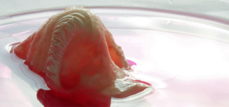
Brain Organoids
Image Source: Yun Li and Julien Muffat

In the study, deletion of the PTEN gene increased activation of the PI3K-AKT pathway and thereby enhanced AKT activity in the human NPs comprising the 3D human cerebral organoids; it promoted cell cycle re-entry and transiently delayed neuronal differentiation, resulting in a marked expansion of the radial glia and intermediate progenitor population. Validating the molecular mechanism at work with PTEN, the investigators used pharmacological AKT inhibitors to reverse the effect of the PTEN deletion. They also found that they could regulate the degree of expansion and folding by tuning the strength of AKT signaling—with reduced signaling resulting in smaller and smooth organoids, and increased signaling producing larger and more folded organoids.

Finally, the researchers utilized the 3D human cerebral organoid system to show that infection with Zika virus impairs cortical growth and folding. In the organoids, Zika infection at the onset of surface folding (day 19 of development) led to widespread apoptosis; and, ten days later, it had severely hampered organoid growth and surface folding. Zika infection of 4-week-old organoids, showed that PTEN mutant organoids were much more susceptible to infection than normal control organoids; notably, they showed increased apoptosis and decreased proliferation of progenitor cells.

“Although not an original goal of our study, we have demonstrated that 3D human cortical organoids can be very effective for Zika modeling—better enabling researchers to observe how human brain tissue reacts to the infection and to test potential treatments,” Li says.


SOURCE  Whitehead Institute via Newswise


By  33rd SquareEmbed


Wednesday, August 28, 2013

human brain grown in a dish

 
Brain Research
Scientists in Austria have grown a three-dimensional, self-organizing model of a developing human brain in the lab using stem cells. The mini-brain may help research brain diseases and treatments more effectively.  


Researchers at the Institute of Molecular Biotechnology in Vienna, Austria, have grown a three-dimensional, self-organizing model of a developing human brain in the lab using stem cells.

The system could be used to model neurological diseases and test treatments in an actual human brain, instead of an animal model that may not develop in exactly the same way, or in human patients.

The cerebral organoid, as the researchers have called it, resembles the early developing regions of a human brain, with distinct regions like the dorsal cortex, the ventral forebrain and even an immature retina.

This is the most complex in vitro human brain tissue created so far. It has the beginning signs of cortical layers, though it can't develop the full complexity of a six-layer human cortex.

Related articles
The scientists were able to grow their organoids from both embryonic stem cells and the less-controversial induced pluripotent stem cells, which can be derived from the skin or blood cells of adult humans. Most of the organoids grew to about 3 or 4 millimeters, about the size of an embryonic human brain at roughly nine weeks, and could survive up to a year in a spinning bioreactor (to circulate nutrients and oxygen).

Though it looks very similar to early-development brain tissue and has active neurons, the organization isn't quite the same as in naturally developing tissue.

Using imaging techniques, the researchers were even able to detect neural activity (see video, above), although this doesn't mean the brain is conscious in anyway.

"The parts are correctly organized, but not put together," Jürgen Knoblich who coordinated the study, explained in a press conference. He describes it as "a car where you have an engine, you have the wheels--but the engine is on the roof…that car would never drive, but you could still take that car and analyze how an engine works."

The stem-cell-derived organoid (right) compared to a developing mouse brain (left)
 Image Source: Marko Repic and Madeline A. Lancaster) 

Scientists have been able to grow other organ tissue in the lab with stem cells, like livers and heart tissue. Unlike with other lab-grown tissue, though, synthetic brain transplants or patches aren't really on the horizon here. The brain is just too complex, for one, and the lack of circulatory system makes it difficult to get enough nutrients and oxygen to the organoid tissue to grow it any larger than 4 millimeters.

Even though they're not exactly put together like human brains, cerebral organoids could be used to analyze diseases like microcephaly, a neurodevelopmental disorder that results in severely small brains.

"Recent work has shown development of human brain is very fundamentally different from the development of the mouse brain," Knoblich said, and certain diseases like microcephaly have been hard to replicate in mice. "Our system is very useful for us as developmental biologists. It allows us to study the human-specific features of brain development." Eventually he would like to be able to model disorders like schizophrenia or autism.


SOURCE  New Scientist

By 33rd SquareSubscribe to 33rd Square