bloc 33rd Square Business Tools - stem cells 33rd Square Business Tools: stem cells - All Post
Showing posts with label stem cells. Show all posts

Wednesday, August 16, 2017

Hard-to-Believe Device Heals Organs with a Single Touch


Researchers have developed a device that can switch cell function to rescue failing body functions with a single touch. The technology, known as Tissue Nanotransfection (TNT), injects genetic code into skin cells, turning those skin cells into other types of cells required for treating diseased conditions.


Researchers at The Ohio State University Wexner Medical Center and Ohio State’s College of Engineering have developed a new technology, Tissue Nanotransfection (TNT), that can generate any cell type of interest for treatment within the patient’s own body. This technology may be used to repair injured tissue or restore function of aging tissue, including organs, blood vessels and nerve cells.

In a new study published in Nature Nanotechnology, first author Daniel Gallego-Perez of Ohio State demonstrated that the technique worked with up to 98 percent efficiently.

"It takes just a fraction of a second. You simply touch the chip to the wounded area, then remove it."
“By using our novel nanochip technology, injured or compromised organs can be replaced. We have shown that skin is a fertile land where we can grow the elements of any organ that is declining,” said Dr. Chandan Sen, director of Ohio State’s Center for Regenerative Medicine & Cell Based Therapies, who co-led the study with L. James Lee, professor of chemical and biomolecular engineering with Ohio State’s College of Engineering in collaboration with Ohio State’s Nanoscale Science and Engineering Center.

Researchers studied mice and pigs in these experiments. In the study, researchers were able to reprogram skin cells to become vascular cells in badly injured legs that lacked blood flow. Within one week, active blood vessels appeared in the injured leg, and by the second week, the leg was saved. In lab tests, this technology was also shown to reprogram skin cells in the live body into nerve cells that were injected into brain-injured mice to help them recover from stroke.

“It takes just a fraction of a second. You simply touch the chip to the wounded area, then remove it,” said Chandan Sen, PhD, director of the Center for Regenerative Medicine and Cell-Based Therapies at The Ohio State University Wexner Medical Center. “At that point, the cell reprogramming begins.”

In a series of lab tests, researchers applied the chip to the injured legs of mice that vascular scans showed had little to no blood flow. “We reprogrammed their skin cells to become vascular cells,” Sen said. “Within a week we began noticing the transformation.” By the second week, active blood vessels had formed, and by the third week, the legs of the mice were saved—with no other form of treatment.

“It extends the concept known as gene therapy, and it has been around for quite some time,” said study collaborator James Lee, PhD, a professor of chemical and biomolecular engineering at Ohio State. “The difference with our technology is how we deliver the DNA into the cells.”

The nanochip, loaded with specific genetic code or certain proteins, is placed on the skin, and a small electrical current creates channels in the tissue. The DNA or RNA is injected into those channels where it takes root and begins to reprogram the cells.

Related articles
“What’s even more exciting is that it not only works on the skin, but on any type of tissue,” Sen said. In fact, researchers were able to grow brain cells on the skin surface of a mouse, harvest them, then inject them into the mouse’s injured brain. Just a few weeks after having a stroke, brain function in the mouse was restored, and it was healed. Because the technique uses a patient’s own cells and does not rely on medication, researchers expect it to be approved for human trials within a year.

“This is difficult to imagine, but it is achievable, successfully working about 98 percent of the time. With this technology, we can convert skin cells into elements of any organ with just one touch. This process only takes less than a second and is non-invasive, and then you're off. The chip does not stay with you, and the reprogramming of the cell starts. Our technology keeps the cells in the body under immune surveillance, so immune suppression is not necessary,” said Sen, who also is executive director of Ohio State’s Comprehensive Wound Center.

TNT technology has two major components: First is a nanotechnology-based chip designed to deliver cargo to adult cells in the live body. Second is the design of specific biological cargo for cell conversion. This cargo, when delivered using the chip, converts an adult cell from one type to another, said first author Daniel Gallego-Perez, an assistant professor of biomedical engineering and general surgery who also was a postdoctoral researcher in both Sen’s and Lee’s laboratories.

TNT doesn’t require any laboratory-based procedures and may be implemented at the point of care. The procedure is also non-invasive. The cargo is delivered by zapping the device with a small electrical charge that’s barely felt by the patient.

“The concept is very simple,” Lee said. “As a matter of fact, we were even surprised how it worked so well. In my lab, we have ongoing research trying to understand the mechanism and do even better. So, this is the beginning, more to come.”


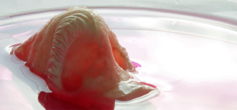
SOURCE  Ohio State University


By  33rd SquareEmbed


Wednesday, July 26, 2017

Stem Cells in the Brain Found to Control Aging


Researchers have found that stem cells in the brain’s hypothalamus govern how fast aging occurs in the body. The finding, made in mice, could lead to new strategies for warding off age-related diseases and extending lifespan. 


In the brain, the hypothalamus is known to regulate important processes including growth, development, reproduction and metabolism. In a 2013 Nature paper, scientists made the surprising finding that the hypothalamus also regulates aging throughout the body.

Now, the researchers at the Albert Einstein College of Medicine have precisely identified the cells in the hypothalamus that control aging: a tiny population of adult neural stem cells, which were known to be responsible for forming new brain neurons.

The team's work has been published in the journal Nature.

"Our research shows that the number of hypothalamic neural stem cells naturally declines over the life of the animal, and this decline accelerates aging."
"Our research shows that the number of hypothalamic neural stem cells naturally declines over the life of the animal, and this decline accelerates aging," says senior author Dongsheng Cai, M.D., Ph.D., professor of molecular pharmacology at the school. "But we also found that the effects of this loss are not irreversible. By replenishing these stem cells or the molecules they produce, it’s possible to slow and even reverse various aspects of aging throughout the body."

The researchers first looked at the fate of those cells as healthy mice got older to see if stem cells in the hypothalamus held the key to aging. The number of hypothalamic stem cells began to diminish when the animals reached about 10 months, which is several months before the usual signs of aging start appearing. "By old age—about two years of age in mice—most of those cells were gone," says Dr. Cai.

Dongsheng Cai

Next, the researchers wanted to learn whether this progressive loss of stem cells was actually causing aging and was not just associated with it. They observed what happened when they selectively disrupted the hypothalamic stem cells in middle-aged mice. "This disruption greatly accelerated aging compared with control mice, and those animals with disrupted stem cells died earlier than normal," says Dr. Cai.

Related articles
The team hypothesized that adding stem cells to the hypothalamus could counteract aging. To test that experiment, the researchers injected hypothalamic stem cells into the brains of middle-aged mice whose stem cells had been destroyed as well as into the brains of normal old mice. In both groups of animals, the treatment slowed or reversed various measures of aging.

Dr. Cai and his colleagues found that the hypothalamic stem cells appear to exert their anti-aging effects by releasing molecules called microRNAs (miRNAs). They are not involved in protein synthesis but instead play key roles in regulating gene expression. miRNAs are packaged inside tiny particles called exosomes, which hypothalamic stem cells release into the cerebrospinal fluid of mice.

The researchers extracted miRNA-containing exosomes from hypothalamic stem cells and injected them into the cerebrospinal fluid of two groups of mice: middle-aged mice whose hypothalamic stem cells had been destroyed and normal middle-aged mice.

This treatment significantly slowed aging in both groups of animals as measured by tissue analysis and behavioral testing that involved assessing changes in the animals’ muscle endurance, coordination, social behavior and cognitive ability.

Dr. Cai and his team are now trying to identify the particular populations of microRNAs and perhaps other factors secreted by these stem cells that are responsible for these anti-aging effects—a first step toward possibly slowing the aging process and treating age-related diseases.

SOURCE  Albert Einstein College of Medicine


By  33rd SquareEmbed


Sunday, January 1, 2017

Researchers Engineer Gene Pathway to Grow Brain Organoids with Surface Folding


Stem Cells

Scientists have demonstrated that 3D human cerebral organoids can be effective in modeling the molecular, cellular, and anatomical processes of human brain development. They also suggest their work could be a new path for identifying the cells affected by Zika virus.


In newly published research in the journal Cell Stem Cell, researchers at Whitehead Institute have found a specific gene pathway that appears to regulate the growth, structure, and organization of the human cortex. They demonstrated that 3D human cerebral organoids—miniature, lab-grown versions of specific brain structures—can be effective in modeling the molecular, cellular, and anatomical processes of human brain development. The researchers suggest their work could also provide a new path for identifying the cells affected by Zika virus.

“We found that increased proliferation of neural progenitor cells (NPs) induces expansion of cortical tissue and cortical folding in human cerebral organoids,” says Yun Li, a lead author of study and post-doctoral researcher at Whitehead Institute. “Further, we determined that deleting the PTEN gene allows increased growth factor signaling in the cell, unleashing its growth potential, and stimulating proliferation.”

Researchers Engineer Gene Pathway to Grow Brain Organoids with Surface Folding

Related articles
The findings lend support to the notion that an increase in the proliferative potential of NPs contributes to the expansion of the human cerebral neocortex, and the emergence of surface folding.

With normal NPs, the human organoid developed into relatively small cell clusters with smooth surface appearance, displaying some features of very early development of a human cortex. However, deleting PTEN allowed the progenitor population to continue expanding and delayed their differentiation into specific kinds of neurons—both key features of the developing human cortex.

“Because the PTEN mutant NPs experienced more rounds of division and retained their progenitor state for an extended period, the organoids grew significantly larger and had substantially folded cortical tissue,” explains Julien Muffat, also a lead author and post-doctoral researcher at MIT's Whitehead Institute.

"We have demonstrated that 3D human cortical organoids can be very effective for Zika modeling."
The researchers found that while PTEN deletion in mouse cells does create a somewhat larger than normal organoid, it does not lead to significant NP expansion or to folding. “Previous studies have suggested that abnormal variation in PTEN expression may play an important role in driving brain development conditions leading to syndromes such as Autism Spectrum Disorders,” says Rudolf Jaenisch, Founding Member of Whitehead Institute and senior author of the study. “Our findings suggest that the PTEN pathway is also an important mechanism for controlling brain-structure differences observed between species.”

Brain Organoids
Image Source: Yun Li and Julien Muffat

In the study, deletion of the PTEN gene increased activation of the PI3K-AKT pathway and thereby enhanced AKT activity in the human NPs comprising the 3D human cerebral organoids; it promoted cell cycle re-entry and transiently delayed neuronal differentiation, resulting in a marked expansion of the radial glia and intermediate progenitor population. Validating the molecular mechanism at work with PTEN, the investigators used pharmacological AKT inhibitors to reverse the effect of the PTEN deletion. They also found that they could regulate the degree of expansion and folding by tuning the strength of AKT signaling—with reduced signaling resulting in smaller and smooth organoids, and increased signaling producing larger and more folded organoids.

Finally, the researchers utilized the 3D human cerebral organoid system to show that infection with Zika virus impairs cortical growth and folding. In the organoids, Zika infection at the onset of surface folding (day 19 of development) led to widespread apoptosis; and, ten days later, it had severely hampered organoid growth and surface folding. Zika infection of 4-week-old organoids, showed that PTEN mutant organoids were much more susceptible to infection than normal control organoids; notably, they showed increased apoptosis and decreased proliferation of progenitor cells.

“Although not an original goal of our study, we have demonstrated that 3D human cortical organoids can be very effective for Zika modeling—better enabling researchers to observe how human brain tissue reacts to the infection and to test potential treatments,” Li says.


SOURCE  Whitehead Institute via Newswise


By  33rd SquareEmbed


Thursday, October 13, 2016

Fully Grown Beating Human Heart Created With Stem Cells

Regenerative Medicine

Scientists have taken some initial steps toward the creation of bioengineered human hearts using donor hearts stripped of cells and induced pluripotent stem cells. The work could lead to custom grown transplantable organs with near zero rejection rates in the future.


Researchers at Massachusetts General Hospital have taken the initial steps toward the creation of bioengineered human hearts using induced pluripotent stem cells (iPSCs) and donor hearts stripped of the elements that would generate an immune response. The work, which includes developing an automated bioreactor system capable of supporting a whole human heart during the recellularization process, was published in the journal Circulation Research.

The scientists are currently developing new approaches to recellularize heart scaffolds to facilitate this regeneration. They are designing and validating novel whole-heart bioreactors that recapitulate cardiac physiology to aid in the maturation of recellularized cardiac matrices.

This project draws from expertise in stem cell biology, developmental biology, physiology, cardiology, and biomedical engineering in pursuit of creating a bioartificial heart that will one day provide a viable treatment option for patients in need of heart transplants.

"Generating functional cardiac tissue involves meeting several challenges," says Jacques Guyette, PhD, of the MGH Center for Regenerative Medicine (CRM), and lead author of the report. "These include providing a structural scaffold that is able to support cardiac function, a supply of specialized cardiac cells, and a supportive environment in which cells can repopulate the scaffold to form mature tissue capable of handling complex cardiac functions."

"Regenerating a whole heart is most certainly a long-term goal that is several years away."
Related articles
The study included 73 human hearts that had been donated through the New England Organ Bank, found to be unsuitable for transplantation and recovered under research consent. Using a scaled-up version of the process originally developed in rat hearts, the team decellularized hearts from both brain-dead donors and from those who had undergone cardiac death. Detailed characterization of the remaining cardiac scaffolds confirmed a high retention of matrix proteins and structure free of cardiac cells, the preservation of coronary vascular and microvascular structures, as well as freedom from human leukocyte antigens that could induce rejection. There was little difference between the reactions of organs from the two donor groups to the complex decellularization process.

Once treated, the organs were mounted for 14 days in an automated bioreactor system developed by the MGH team that both perfused the organ with nutrient solution and applied environmental stressors such as ventricular pressure to reproduce conditions within a living heart. Analysis of the regenerated tissue found dense regions of iPSC-derived cells that had the appearance of immature cardiac muscle tissue and demonstrated functional contraction in response to electrical stimulation.

"Regenerating a whole heart is most certainly a long-term goal that is several years away, so we are currently working on engineering a functional myocardial patch that could replace cardiac tissue damaged due a heart attack or heart failure," says Guyette.

"Among the next steps that we are pursuing are improving methods to generate even more cardiac cells - recellularizing a whole heart would take tens of billions -- optimizing bioreactor-based culture techniques to improve the maturation and function of engineered cardiac tissue, and electronically integrating regenerated tissue to function within the recipient's heart."

Team leader Harald Ott, MD, of the MGH CRM and the Department of Surgery, , an assistant professor of Surgery at Harvard Medical School, adds, "Generating personalized functional myocardium from patient-derived cells is an important step towards novel device-engineering strategies and will potentially enable patient-specific disease modeling and therapeutic discovery. Our team is excited to further develop both of these strategies in future projects."


SOURCE  EurekAlert


By  33rd SquareEmbed


Friday, September 16, 2016

Experimental Regeneraitve Medicine Treatment Returns Use of Arms and Hands to Paralyzed Man


Regenerative Medicine

Using pioneering stem cell surgery, doctors have made excellent progress in returning function to a young man recently paralyzed in a traffic accident. This is he latest example of how the emerging field of regenerative medicine may have the potential to improve the lives of thousands of patients who have suffered a severe spinal cord injury.


Doctors at Keck Medical Center at the University of Southern California (USC) have become the first in California to inject an experimental treatment made from stem cells, AST-OPC1, into the damaged cervical spine of a recently paralyzed 21-year-old man as part of a multi-center clinical trial.

The breakthrough surgery is the latest example of how the emerging field regenerative medicine may have the potential to improve the lives of thousands of patients who have suffered a severe spinal cord injury.

This spring, just before his 21st birthday, Kristopher (Kris) Boesen of Bakersfield suffered a traumatic injury to his cervical spine when his car fishtailed on a wet road, hit a tree and slammed into a telephone pole.His parents Rodney and Annette Boesen were warned there was a good chance their son would be permanently paralyzed from the neck down. However, they also learned that Kris could possibly qualify for a clinical study that might help.

Charles Liu

Leading the surgical team and working in collaboration with Rancho Los Amigos National Rehabilitation Center and Keck Medicine of USC, Charles Liu, MD, PhD, director of the USC Neurorestoration Center, injected an experimental dose of 10 million AST-OPC1 cells directly into Kris’ cervical spinal cord in early April.

"With this study, we are testing a procedure that may improve neurological function, which could mean the difference between being permanently paralyzed and being able to use one’s arms and hand."
“Typically, spinal cord injury patients undergo surgery that stabilizes the spine but generally does very little to restore motor or sensory function,” explains Liu. “With this study, we are testing a procedure that may improve neurological function, which could mean the difference between being permanently paralyzed and being able to use one’s arms and hands. Restoring that level of function could significantly improve the daily lives of patients with severe spinal injuries.”

Related articles
Kris began to show signs of improvement as early as two weeks after the surgery. Three months later, he’s able to feed himself, use his cell phone, write his name, operate a motorized wheelchair and hug his friends and family. Improved sensation and movement in both arms and hands also makes it easier for Kris to care for himself, and to envision a life lived more independently.

“As of 90 days post-treatment, Kris has gained significant improvement in his motor function, up to two spinal cord levels,” said Dr. Liu. “In Kris’ case, two spinal cord levels means the difference between using your hands to brush your teeth, operate a computer or do other things you wouldn’t otherwise be able to do, so having this level of functional independence cannot be overstated.”

“All I’ve wanted from the beginning was a fighting chance,” said Kris, who has a passion for fixing up and driving sports cars and was studying to become a life insurance broker at the time of the accident. “But if there’s a chance for me to walk again, then heck yeah! I want to do anything possible to do that.”

The stem cell procedure Kris underwent is part of a Phase 1/2a clinical trial that is evaluating the safety and efficacy of escalating doses of AST-OPC1 cells developed by Fremont, California-based Asterias Biotherapeutics. AST-OPC1 cells are made from embryonic stem cells by carefully converting them into oligodendrocyte progenitor cells (OPCs), which are cells found in the brain and spinal cord that support the healthy functioning of nerve cells. In previous laboratory studies, AST-OPC1 was shown to produce neurotrophic factors, stimulate vascularization and induce remyelination of denuded axons.

All are critical factors in the survival, regrowth and conduction of nerve impulses through axons at the injury site, according to Edward D. Wirth III, MD, PhD, chief medical director of Asterias and lead investigator of the study, named SCiStar.

The team is currently looking for more clinical subjects for the study.

“At the 10 million cell level, we’re now in a dose range that is the human equivalent of where we were when we saw efficacy in pre-clinical studies,” says Wirth. “While we continue to evaluate safety first and foremost, we are also now looking at how well treatment might help restore movement in these patients.”


SOURCE  USC Stem Cell


By  33rd SquareEmbed


Saturday, April 30, 2016

Substance Related to Vitamin B3 May Hold Key to Regenerative Medicine


Regenerative Medicine

Researchers have discovered that the substance nicotinamide riboside (NR) may have regenerative effects, having used it to prolong the lives of elderly mice. NR helps stimulate mitochondria to help the body correct damage, a major accumulating factor in aging.


Nicotinamide riboside (NR) has already been shown in several studies to be effective in boosting metabolism. The molecule is a pyridine-nucleoside form of vitamin B3 that functions as a precursor to nicotinamide adenine dinucleotide or NAD+. David Sinclair published a study in 2013 showing NAD+ levels decrease with age in mice, making NR a key research focus for life extension and regenerative medicine.

Now a team of researchers at EPFL's Laboratory of Integrated Systems Physiology (LISP), have unlocked even more of NR's secrets.

Related articles
In an article recently published recently in the journal Science  the researchers describe the positive effects of NR on the functioning of stem cells.

These effects can only be described as restorative.

"We demonstrated that fatigue in stem cells was one of the main causes of poor regeneration or even degeneration in certain tissues or organs."
Mice, like all mammals, age, the regenerative capacity of certain organs (such as the liver and kidneys) and muscles (including the heart) diminishes. Their ability to repair them following an injury is also affected. This leads to many of the disorders typical of aging.

Researchers all over the world have been hunting for restorative tools including technological ones. For instance Bruce Eaton has been spearheading the creation of technology-based tools for anti-aging pharmaceutical drug discovery. The new research could make it possible to eventually take a supplement that could forestall the advent of aging.

In the new study, PhD candidate Hongbo Zhang wanted to understand how the regeneration process deteriorated with age. To do this, he teamed up with colleagues from ETH Zurich, the University of Zurich and universities in Canada and Brazil. Using several markers, Zhang was able to identify the molecular chain that regulates how mitochondria - the "powerhouse" of the cell - function and how they change with age. The role that mitochondria play in metabolism has already been substantially researched, "but we were able to show for the first time that their ability to function properly was important for stem cells," said LISP head Johan Auwerx.

Normally in young bodies stem cells regenerate damaged organs by producing new specific cells, reacting to signals sent by the body. "We demonstrated that fatigue in stem cells was one of the main causes of poor regeneration or even degeneration in certain tissues or organs," said Zhang.

In the study the researchers targeted the molecules that help the mitochondria to function properly. "We gave nicotinamide riboside to 2-year-old mice, which is an advanced age for them," said Zhang. "This substance, which is close to vitamin B3, is a precursor of NAD+, a molecule that plays a key role in mitochondrial activity. And our results are extremely promising: muscular regeneration is much better in mice that received NR, and they lived longer than the mice that didn't get it."

Vitamin B3 in the form of Niacin has long believed to have other anti-aging benefits. There's good evidence that it helps reduce atherosclerosis, or hardening of the arteries in some people. For people who have already had a heart attack, niacin seems to lower the risk of a second one. In addition, niacin is an FDA-approved treatment for pellagra, a rare condition that develops from niacin deficiency.

Niacin has also been studied as a treatment for many other health problems. There's some evidence that it might help lower the risk of Alzheimer's disease, cataracts, osteoarthritis, and type 1 diabetes.

Niacin helps in creating sex hormones for people suffering through sexual disorders like impotence and erectile dysfunction.

Niacin occurs naturally in many foods, including greens, meat, poultry, fish, and eggs, though in a fraction of the dose shown to achieve changes in cholesterol. Niacin is also commonly used as a food additive, to increase the nutritional value of manufactured foodstuff.

"This work could have very important implications in the field of regenerative medicine," said Auwerx. "We are not talking about introducing foreign substances into the body but rather restoring the body's ability to repair itself with a product that can be taken with food." This work on the aging process also has potential for other areas of aging and disease.

So far, no negative side effects have been observed following the use of Nicotinamide riboside, even at high doses. But caution many more studies are required.

SOURCE  Medical Xpress


By 33rd SquareEmbed


Monday, February 15, 2016

The Chief Medical Officer at Human Longevity Says We Will Be Celebrating our 200th Birthdays


Anti-Aging

Human Longevity Inc.'s Brad Perkins says technological innovations including cloud-based systems could spark a medical revolution in tackling age-related diseases.


Brad Perkins is a visionary physician, scientist, and executive who is responsible for leading all clinical and therapeutic operations at the Human Longevity Inc. This includes collecting and utilizing phenotype data, development of the consumer clinics business, and guiding stem cell therapeutics.

"With machine learning, scientists can connect medical genome to sequence data and, therefore, understand the fundamental causes of aging."
According to Perkins, technological innovations including cloud-based systems could spark a medical revolution in tackling age-related diseases.

Research into diseases such as Alzheimer's, diabetes and cancer could be advanced by new technology and could potentially see the human lifespan eventually double, states Perkins, who spoke at this year's World Government Summit in Dubai.

Technology cited included integrated data bases, cloud-based systems, advanced clinical imaging and machine, which could help improve knowledge of the human genome.

“With machine learning, scientists can connect medical genome to sequence data and, therefore, understand the fundamental causes of aging. By comparing physiological to chronological age and building direct medical intervention methods for age, we are closer to preventing age related diseases.”

Related articles
Prior to joining HLI, Dr. Perkins was Executive Vice President for Strategy and Innovation, and Chief Transformation Officer at Vanguard Health Systems, a large multi-state, for-profit, integrated health services provider with nearly 46,000 employees. He helped transform Vanguard from a traditional fee for service healthcare model, to a fee for value, “population health” model. Some of his innovative solutions there included: establishing Accountable Care Organizations to improve primary care, implementing an award winning tele-radiology program, and starting a $167 million venture capital fund to support Vanguard’s transformation programs.

Dr. Perkins began his career at the Centers for Disease Control and Prevention (CDC) in 1989 after completing his residency training and chief residency in internal medicine at Baylor College of Medicine. At the CDC he led some of the most important and high profile programs and published more than 120 peer-reviewed publications and book chapters.

He first joined and then led the Meningitis and Special Pathogens Branch where he investigated global bacterial disease epidemics. He co-discovered the bacteria which causes Cat Scratch Diseases and conducted translational research leading to development of several new bacterial meningitis and pneumonia vaccines. In 2001 Dr. Perkins led the investigations into the anthrax attacks in the United States, the largest and highest profile investigation ever conducted by CDC. In 2005 he was appointed CDC’s Chief Strategy and Innovation Officer, a position in which he managed a $11.2 billion budget, and 15,000 employees with offices in more than 50 countries. Working closely with the CDC Director, he built a $2 billion state-of-the-art emergency response capability and positioned the improvement of population health as a focus of the healthcare reform movement within the White House administration at that time.

Dr. Perkins is a member of the RAND Health Board, and he is the chairman of the advisory board for Esther Dyson’s nonprofit, HICCup, sponsor of the “Way to Wellville” community health competition. He received his BA in Microbiology and his MD from the University of Missouri-Columbia, and an MBA from Emory University. He is Board Certified in Internal Medicine.


SOURCE  World Government Summit


By 33rd SquareEmbed


Saturday, January 23, 2016

Algorithm Developed to Help Convert Cells from One Type to Another


Regenerative Medicine

Researchers have develop an algorithm that takes the field of cell reprogramming forward, by helping to pinpoint potential errors and cancerous mutations when cells are changed from one type to another. The technique should help to advance the field of regenerative medicine dramatically.

Researchers from the Duke-NUS Medical School (Duke-NUS), the University of Bristol, Monash University and RIKEN have created an algorithm that can predict the factors required to convert one human cell type to another. The findings were recently published in the journal Nature Genetics, have significant implications for regenerative medicine and lay the groundwork for further research into cell reprogramming.

Related articles
Cell types in the body are not fixed; cell type can be reprogrammed, or converted, to become another cell type by the addition of a unique set of cellular factors. This process was established by Shinya Yamanaka, in Nobel prize-winning work involved the reprogramming of fibroblast cells from the skin to induced pluripotent stem cells (iPS).

Theoretically, stem cells can be reprogrammed for use in regenerative medicine techniques. To date the practice has not been perfected though. There are technical and safety concerns in converting cells because of  the accumulation of unpredictable errors, including cancerous mutations in the reprogrammed cells.

Despite this development, determining the unique set of cellular factors that is needed to be manipulated for each cell conversion is a long and costly process that involved much trial and error. As a result, this first step of identifying the key set of cellular factors for cell conversion is the major obstacle researchers and doctors face in the field of cell reprogramming.

The researchers worked for five years to develop a computational algorithm to predict the cellular factors for cell conversions. The algorithm, called Mogrify(1), is able to predict the optimal set of cellular factors required for any given cell conversion.

Mogrify cell reprogramming


Mogrify uses a network-based algorithm designed to find transcription factors that impart the most influence on changes in cellular state. This website will allow you to explore possible reprogramming experiments, different collections of transcription factors as well as the look at the changes in the regulatory network.

"One of the first clinical applications that we hope to achieve with this innovative approach would be to reprogram 'defective' cells from patients into 'functioning' healthy cells."
"Mogrify acts like a 'world atlas' for the cell and allows us to map out new territories in cell conversions in humans," explained Dr Rackham, who is from the Systems Genetics of Complex Disease Laboratory at Duke-NUS. "One of the first clinical applications that we hope to achieve with this innovative approach would be to reprogram 'defective' cells from patients into 'functioning' healthy cells, without the intermediate iPS step. These then can be re-implanted into patients, and should, in practice, effectively enable new regenerative medicine techniques."

Associate Professor Enrico Petretto, co-author of the study and head of the Systems Genetics of Complex Disease Laboratory in the Centre for Computational Biology at Duke-NUS, highlighted that since Mogrify is completely data-driven, its robustness and accuracy can only continue to improve as more comprehensive data are collected and input into the framework.

"Mogrify is a game-changing method that leverages big-data and systems-biology; this will inspire new translational applications as the result of the work and expertise here at Duke-NUS," said Assoc Prof Petretto.

Mogrify is available online for other researchers and scientists. The team at Duke-NUS now plan to focus on Mogrify's application in translational medicine. Collaborative efforts between research groups within Duke-NUS are already in place to apply the algorithm to help develop treatments for specific diseases, such as cancer.


SOURCE  EurekAlert


By 33rd SquareEmbed


Wednesday, January 20, 2016

New Initiative Aims to Put Canada at the Forefront of Regenerative Medicine


Regenerative Medicine

The Canadian government has announced a funding kick start for the new Centre for Commercialization of Regenerative Medicine to help establish a stem-cell therapy development facility. The new center aims to accelerate the development and adoption of cell manufacturing biotechnologies.


The newly-elected government in Canada has announced they will provide $20 million start-up capital for the Centre for Commercialization of Regenerative Medicine (CCRM) to help
establish a stem-cell therapy development facility in Toronto.

The Centre for Advanced Therapeutic Cell Technologies will be the first such facility in the world to use a collaborative approach between research institutions and industry to solve cell therapy manufacturing challenges, according to the government.

Related articles
"Regenerative medicine is the future and not only is it the future, it's a branch of medicine that Canada and the province of Ontario are actually quite good at."
"Regenerative medicine is the future and not only is it the future, it's a branch of medicine that Canada and the province of Ontario are actually quite good at," said Prime Minister Justin Trudeau. "The medical advances and innovations happening right here in Toronto are world class."

Trudeau made the announcement this morning on an empty floor of the MaRS tower in downtown Toronto, designed for medical and research labs, where the stem cell facility will be located.

The stem cell facility is government funded by some money is also coming from GE Healthcare. The federal money will be provided once certain terms and conditions are met, the government said.

That money will be used to "support improvements to the new facility and the purchase of specialized equipment."

CCRM president Michael May said Canada is leading the charge as the industry moves toward mass production of stem cells and cell-based products.

“The industry and the science has evolved, and we’ve been leading for 60 years, the science of regenerative medicine and stem cells, it’s now time to lead the industrialization and commercialization,” May told CTV’s Canada AM.

The global market for cell-based therapies is expected to surpass the $20 billion USD mark by 2025, with an annual growth rate of 21%. The main targets for cell-based therapies are high impact disease areas with significant unmet need, including cancer, heart disease, neurodegenerative diseases, musculoskeletal disorder and autoimmune diseases.


SOURCE  CBC


By 33rd SquareEmbed


Thursday, June 4, 2015

One Secret to a Longer Life—Be a Female

Aging
Human supercentenarians share at least one thing in common--over 95 percent are women. Scientists have long observed differences between the sexes when it comes to aging, but there is no clear explanation for why females live longer. 


Human supercentenarians share at least one trait in common—over 95 percent are women. Scientists have long noticed the differences between the sexes in aging, but there is still no clear explanation for why females live longer.

Related articles
Now, in a discussion paper of what we know about stem cell behavior and sex, Stanford University researchers Ben Dulken and Anne Brunet argue that it's time to look at differences in regenerative decline between men and women. This line of research could open up new explanations for how the sex hormones estrogen and testosterone, or other factors, affect lifespan.

It's known that estrogen has direct effects on stem cell populations in female mice, from increasing the number of blood stem cells (which is very helpful during pregnancy) to enhancing the regenerative capacity of brain stem cells at the height of estrus. Whether these changes have a direct impact on lifespan is what's yet to be explored. Recent studies have already found that estrogen supplements increase the lifespan of male mice, and that human eunuchs live about 14 years longer than non-castrated males.

the effect of sex on the aging of stem cells

"As the search continues for ways to ameliorate the aging process and maintain the regenerative capacity of stem cells, let us not forget one of the most effective aging modifiers: sex."


Though interactions between stem cells, aging, and sex have been topics of great interest, the intersection of all three—the effect of sex on the aging of stem cells—has not been well studied. However, several mechanisms could be involved in establishing and perpetuating sexual dimorphism during the aging of stem cells.

More work is also needed to understand how genetics impacts stem cell aging between the sexes. Scientists have seen that knocking out different genes in mice can add longevity benefits to one sex but not the other, and that males in twin studies have shorter telomeres—a sign of shorter cellular lifespan—compared to females.

"It is likely that sex plays a role in defining both lifespan and healthspan, and the effects of sex may not be identical for these two variables," the authors write. "As the search continues for ways to ameliorate the aging process and maintain the regenerative capacity of stem cells, let us not forget one of the most effective aging modifiers: sex."

SOURCE  EurekAlert

By 33rd SquareEmbed

Tuesday, May 19, 2015

Drug Candidate Seems To Wake Up Aging Brains and Muscles

Anti-Aging
Researchers have discovered that a small-molecule drug simultaneously perks up old stem cells in the brains and muscles of mice, a finding that could lead to drug interventions for humans that would make aging tissues throughout the body act young again.


Researchers at UC Berkeley have discovered a small-molecule drug that may be the fountain of youth for aging brains and muscles. In mouse models the drug is working wonders and they hope their findings will lead to a drug that does the same thing for humans.

"We established that you can use a single small molecule to rescue essential function in not only aged brain tissue but aged muscle."


“We established that you can use a single small molecule to rescue essential function in not only aged brain tissue but aged muscle,” said co-author David Schaffer, director of the Berkeley Stem Cell Center and a professor of chemical and biomolecular engineering. “That is good news, because if every tissue had a different molecular mechanism for aging, we wouldn’t be able to have a single intervention that rescues the function of multiple tissues.”

Their findings were published in the journal Oncotarget.

In mice, as in humans, stem cell function declines with age. The drug interferes with the growth factor, TGF-beta1 that keeps those stem cells from regenerating.

Related articles
By inhibiting TGF-beta1, researchers were able to enhance neurogenesis and muscle regeneration in 24-month-old laboratory mice, the age equivalent of 80-year-old humans.

If humans were to respond the same way, age-related degeneration such as loss of agility, mobility, memory, learning and independence could be treatable.

The small-molecule drug is already being used in trials as an anticancer agent. Researchers say it holds a lot of promise when when used on stem cells, because it can regenerate both the mind and the body.
hippocampus
Images of cells in the brain’s
hippocampus show that the growth
 factor TGF-beta1 (stained red)
 is barely present in young tissue
 but ubiquitous in old tissue,
where it suppresses stem
cell regeneration and
contributes to aging.
“You can simultaneously improve tissue repair and maintenance repair in completely different organs, muscle and brain,” said UC scientist Irina Conboy.

“Based on our earlier papers, the TGF-beta1 pathway seemed to be one of the main culprits in multi-tissue aging,” said Conboy, an associate professor of bioengineering. “That one protein, when upregulated, ages multiple stem cells in distinct organs, such as the brain, pancreas, heart and muscle. This is really the first demonstration that we can find a drug that makes the key TGF-beta1 pathway, which is elevated by aging, behave younger, thereby rejuvenating multiple organ systems.”

She and her colleagues caution this new drug “is only a first step toward a therapy, since other biochemical cues also regulate adult stem cell activity.” They noted that this is only a first step toward a therapy, since other biochemical cues also regulate adult stem cell activity. Schaffer and Conboy’s research groups are now collaborating on a multi-pronged approach in which modulation of two key biochemical regulators might lead to safe restoration of stem cell responses in multiple aged and pathological tissues.

“The challenge ahead is to carefully retune the various signaling pathways in the stem cell environment, using a small number of chemicals, so that we end up recalibrating the environment to be youth-like,” Conboy said. “Dosage is going to be the key to rejuvenating the stem cell environment.”


SOURCE  Berkeley Research

By 33rd SquareEmbed