bloc 33rd Square Business Tools - connectome 33rd Square Business Tools: connectome - All Post
Showing posts with label connectome. Show all posts

Wednesday, October 22, 2014


 Neuroscience
Neuroscience researchers have created Golgi, an interactive map of a rat brain that makes exploring the brain as easy as using Google Maps.


What is the link in your brain between smell and memory? Where’s the connection between habits and Parkinson’s disease? How does one detour into addiction?

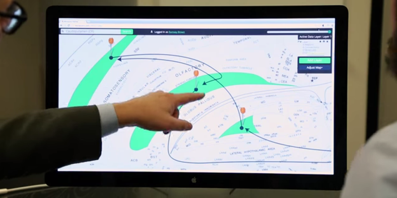
To answer these and other complex scientific and medical questions, two University of Southern California scientists have built Golgi, an interactive map of a rat brain that makes exploring the brain as easy as using Google Maps.

The map can be found online at www.useGolgi.com.

www.useGolgi.com


"We have a big advantage because we’re the only group — really in the world — that has a flat map of the brain."


The new Web app, unveiled today, offers details at the click of a button about how the regions of the brain communicate and interact. Golgi will help accelerate the research and treatment of diseases like Parkinson’s and depression by layering complex scientific data onto a single simple brain map that provides information to doctors and researchers quickly and intuitively.

“We have a big advantage because we’re the only group — really in the world — that has a flat map of the brain,” said Larry Swanson, National Academy member, professor of biological sciences at the USC Dornsife College of Letters, Arts and Sciences and recent past president of the Society for Neuroscience.

Swanson, a longtime pillar of the neuroscience community, collaborated with 25-year-old USC graduate student Ramsay Brown, who designed the program as an undergraduate worker in Swanson’s lab.

Related articles
Golgi takes the existing pool of knowledge about the brain and makes it easy to access by scientists and doctors, saving time and offering potentially new connections. It uses research on rat brains, which are close enough to human brains to offer valuable insights but are far easier to study and therefore represent a larger pool of research data.

To display the brain’s three-dimensional structure on two-dimensional screens, Swanson and Brown used the embryonic brain — which begins as a flat sheet of cells — as a guide. This flattens the brain and keeps related portions of the brain located close together. Flattening the brain lets users click around and display connectome and other data directly on regions they’re interested in learning about for research or treatment.

“We designed a really intuitive way to explore the more nuanced details about the brain and connectome,” Brown said. “Making this data easy and accessible will improve how scientists and doctors explore, explain and treat human conditions and restore quality of life — and that’s really special to us.”

Brown and Swanson think this program is just the beginning.

Connectomics, the subfield of neuroscience that studies and maps the brain’s wiring, is advancing quickly and providing better maps as the technique evolves. Programs like Golgi will help doctors and researchers make sense of these new maps and make better medical and scientific decisions faster.

“Many people now think that understanding these neurological diseases is going to require understanding the circuitry of the brain,” Swanson said.


SOURCE  USC

By 33rd SquareEmbed

Monday, October 20, 2014


 Mind Uploading
Mind uploading is the process of transferring the contents of an individual’s biological mind, to another substrate.  Dr. Ken Hayworth has now outlined a proposal to achieve this Apollo-scale task in a roadmap.


One of the greatest forever unresolved philosophical questions: What is reality? Will get a whole lot more complex if Ken Hayworth’s interpretation of the logical conclusion to his work is attained. Hayworth’s work: brain mapping. The proposed logical conclusion: mind uploading.

Mind uploading is the process of transferring the contents of an individual’s biological mind, including: memories, consciousness and personality traits…to another substrate, such as a computer.

Related articles
There are countless ways human brain mapping will help humanity, notably for understanding and combating neurological disorders. But Hayworth’s ultimate goal of mind uploading pushes the research into the realm of a post-human possibility, a place few scientists have opened their own minds to.

According to Hayworth, "Mind uploading is the logical conclusion of an advanced neuroscience.  If we really understand how the brain works, we will be able to build that brain."

Ken Hayworth

"Mind uploading is the logical conclusion of an advanced neuroscience.  If we really understand how the brain works, we will be able to build that brain."


Hayworth believes that chemical fixation, not cryonics, offers the most promising method for capturing a complete human connectome, which could then be used to recreate a brain digitally. Current methods have only just been able to plasticize a whole mouse brain, but digitizing the connectome is still a huge technological challenge.

Hayworth estimates that in order to capture a human brain, the technology will have to be scaled 100 million-fold. This is a prerequisite to mind uploading.

Mind Uploading

Several large scale scientific endeavors to better understand the brain are underway; their success or failure could set the tone for the future mainstream acceptance or rejection of Hayworth's roadmap to mind uploading. His 'logical conclusion' is still the stuff of science fiction – for now.


SOURCE  Galactic Public Archives

By 33rd SquareEmbed

Wednesday, September 24, 2014


 Neuroscience
Researchers have created models of neurons in the visual cortex of a mouse in order to better understand—and make predictions about—how neurons relay and code visual information.


Scientists at the Allen Institute for Brain Science have created models of neurons in the visual cortex of a mouse in order to better understand—and make predictions about—how neurons relay and code visual information.

This movie shows preliminary data of 10,000 neurons: 8,000 replicated pyramidal cells (an excitatory neuron, shown first) and 2,000 replicated interneurons (an inhibitory neuron, shown second).

10,000 Neurons Modelled


Related articles
Copies of these two cells are displayed one after another in the movie until approximately 10% of the whole model is shown in the video.(More than that would be too cluttered to see what is happening.)

Toward the middle of the movie, the cells are colored according to their properties: excitatory in red and inhibitory in blue. The second half of the movie illustrates activity of the cells in a two second-long simulation. The cells that fire action potentials are intermittently highlighted, giving the viewer an impression of the simulated neuronal activity.

The results in themselves are amazing, but the scale compared with the 80 billion plus neurons in the human brain demonstrate the monumental task computational neuroscience projects like Europe's Human Brain Project and the US BRAIN Initiative have ahead of them.  Completing a connectome of a human brain in silico, will be some years away.


SOURCE  Allen Institute for Brain Science

By 33rd SquareEmbed

Monday, September 8, 2014

Researchers Looking To Map The Brain Find It Even More Complex Than They Thought

 Neuroscience
Researchers have found that the brain's wiring is more complex than expected – one set of neural wires can trigger different reactions, depending on how it fires. The work opens new questions for scientists trying to map the brain's connections.


Scientists at Stanford's Bio-X have raised an entirely new set of questions when they sought answers about connections between two brain regions.

"There's a lot of excitement about being able to make a map of the brain with the idea that if we could figure out how it is all connected we could understand how it works,"  researcher Joanna Mattis said. "It turns out it's so much more dynamic than that."

Mattis is a co-first author on a paper describing the work published recently in the Journal of Neuroscience. Julia Brill, then a postdoctoral scholar, was the other co-first author.

Mattis had been a graduate student in the lab of Karl Deisseroth, professor of bioengineering and of psychiatry and behavioral sciences, where she helped work on a new technique called optogenetics. That technique allows neuroscientists to selectively turn parts of the brain on and off to see what happens. She wanted to use optogenetics to understand the wiring of a part of the brain involved in spatial memory – it's what makes a mental map of your surroundings as you explore a new city, for example.

Related articles
Scientists already knew that when an animal explores habitats, two parts of the brain are involved in the initial exploring phase and then in solidifying a map of the environment – the hippocampus and the septum.

When an animal is exploring an environment, the neurons in the hippocampus fire slow signals to the septum, essentially telling the septum that it's busy acquiring information. Once the animal is done exploring, those same cells fire off intense signals letting the septum know that it's now locking that information into memory. The scientists call this phase consolidation. The septum uses that information to then turn around and regulate other signals going into the hippocampus.

"I wanted to study the hippocampus because on the one hand so much was already known – there was already this baseline of knowledge to work off of. But then the question of how the hippocampus and septum communicate hadn't been accessible before optogenetics," Mattis said.

connectome
Neural wiring diagrams are even more complex than initially speculated according to new research
Neurons in the hippocampus were known to fire in a rhythmic pattern, which is a particular expertise of John Huguenard, a professor of neurology. Mattis obtained an interdisciplinary fellowship through Stanford Bio-X, which allowed her to combine the Deisseroth lab's expertise in optogenetics with the rhythmic brain network expertise of Julia Brill from the Huguenard lab.

"There's a lot of excitement about being able to make a map of the brain with the idea that if we could figure out how it is all connected we could understand how it works."


Mattis and Brill used optogenetics to prompt neurons of the hippocampus to mimic either the slow firing characteristic of information acquisition or the rapid firing characteristic of consolidation. When they mimicked the slow firing they saw a quick reaction by cells in the septum. When they mimicked the fast consolidation firing, they saw a much slower response by completely different cells in the septum.

Same set of wires – different outcome. That's like turning on different lights depending on how hard you flip the switch. "This illustrates how complex the brain is," Mattis said.

This research has raised a whole new set of questions, Mattis said. They more or less understand the faster reaction, but what is causing the slower reaction? How widespread is this phenomenon in the brain?

"The other big picture thing that we opened up but didn't answer is: How can you then tie this back to the circuit overall and learning memory?" Mattis said. "Those would be exciting things to follow up on for future projects."


SOURCE  Stanford

By 33rd SquareEmbed

Thursday, July 3, 2014

CLARITY Improvement Makes It Even Easier To See Inner Workings of the Brain

 Neuroscience
Scientists have improved on their original technique for peering into the intact brain, making it more reliable and safer. The results could help scientists unravel the inner connections of how thoughts, memories or diseases arise.


Last year Karl Deisseroth, a Stanford professor of bioengineering and of psychiatry and behavioral sciences, announced a new way of peering into a brain – removed from the body – that provided spectacular fly-through views of its inner connections. Since then laboratories around the world have begun using the technique, called CLARITY, with some success, to better understand the brain's wiring.

However, Deisseroth said that with two technological fixes CLARITY could be even more broadly adopted. The first problem was that laboratories were not set up to reliably carry out the CLARITY process. Second, the most commonly available microscopy methods were not designed to image the whole transparent brain. "There have been a number of remarkable results described using CLARITY," Deisseroth said, "but we needed to address these two distinct challenges to make the technology easier to use."

In a recent Nature Protocols paper, Deisseroth presented solutions to both of those bottlenecks. "These transform CLARITY, making the overall process much easier and the data collection much faster," he said. He and his co-authors, including postdoctoral fellows Raju Tomer and Li Ye and graduate student Brian Hsueh, anticipate that even more scientists will now be able to take advantage of the technique to better understand the brain at a fundamental level, and also to probe the origins of brain diseases.

"This work shares the spirit of the BRAIN Initiative goal of building new technologies to understand the brain – including the human brain."


This paper may be the first to be published with support of the White House BRAIN Initiative, announced last year with the ambitious goal of mapping the brain's trillions of nerve connections and understanding how signals zip through those interconnected cells to control our thoughts, memories, movement and everything else that makes us us.

"This work shares the spirit of the BRAIN Initiative goal of building new technologies to understand the brain – including the human brain," said Deisseroth, who is also a Stanford Bio-X affiliated faculty member.

Eliminating fat


When you look at the brain, what you see is the fatty outer covering of the nerve cells within, which blocks microscopes from taking images of the intricate connections between deep brain cells. The idea behind CLARITY was to eliminate that fatty covering while keeping the brain intact, complete with all its intricate inner wiring.

The way Deisseroth and his team eliminated the fat was to build a gel within the intact brain that held all the structures and proteins in place. They then used an electric field to pull out the fat layer that had been dissolved in an electrically charged detergent, leaving behind all the brain's structures embedded in the firm water-based gel, or hydrogel. This is called electrophoretic CLARITY.

The electric field aspect was a challenge for some labs. "About half the people who tried it got it working right away," Deisseroth said, "but others had problems with the voltage damaging tissue." Deisseroth said that this kind of challenge is normal when introducing new technologies. When he first introduced optogenetics, which allows scientists to control individual nerves using light, a similar proportion of labs were not initially set up to easily implement the new technology, and ran into challenges.

To help expand the use of CLARITY, the team devised an alternate way of pulling out the fat from the hydrogel-embedded brain – a technique they call passive CLARITY. It takes a little longer, but still removes all the fat, is much easier and does not pose a risk to the tissue. "Electrophoretic CLARITY is important for cases where speed is critical, and for some tissues," said Deisseroth, who is also the D.H. Chen Professor. "But passive CLARITY is a crucial advance for the community, especially for neuroscience." Passive CLARITY requires nothing more than some chemicals, a warm bath and time.

Many groups have begun to apply CLARITY to probe brains donated from people who had diseases like epilepsy or autism, which might have left clues in the brain to help scientists understand and eventually treat the disease. But scientists, including Deisseroth, had been wary of trying electrophoretic CLARTY on these valuable clinical samples with even a very low risk of damage. "It's a rare and precious donated sample, you don't want to have a chance of damage or error," Deisseroth said. "Now the risk issue is addressed, and on top of that you can get the data very rapidly."

Fast CLARITY imaging in color


The second advance had to do this rapidity of data collection. In studying any cells, scientists often make use of probes that will go into the cell or tissue, latch onto a particular molecule, then glow green, blue, yellow or other colors in response to particular wavelengths of light. This is what produces the colorful cellular images that are so common in biology research. Using CLARITY, these colorful structures become visible throughout the entire brain, since no fat remains to block the light.

Related articles
But here's the hitch. Those probes stop working, or get bleached, after they've been exposed to too much light. That's fine if a scientist is just taking a picture of a small cellular structure, which takes little time. But to get a high-resolution image of an entire brain, the whole tissue is bathed in light throughout the time it takes to image it point by point. This approach bleaches out the probes before the entire brain can be imaged at high resolution.

The second advance of the new paper addresses this issue, making it easier to image the entire brain without bleaching the probes. "We can now scan an entire plane at one time instead of a point," Deisseroth said. "That buys you a couple orders of magnitude of time, and also efficiently delivers light only to where the imaging is happening." The technique is called light sheet microscopy and has been around for a while, but previously didn't have high enough resolution to see the fine details of cellular structures. "We advanced traditional light sheet microscopy for CLARITY, and can now see fine wiring structures deep within an intact adult brain," Deisseroth said. His lab built their own microscope, but the procedures are described in the paper, and the key components are commercially available. Additionally, Deisseroth's lab provides free training courses in CLARITY, modeled after his optogenetics courses, to help disseminate the techniques.

Brain imaging to help soldiers


The BRAIN Initiative is being funded through several government agencies including the Defense Advanced Research Projects Agency (DARPA), which funded Deisseroth's work through its new Neuro-FAST program. Deisseroth said that like the National Institute of Mental Health (NIMH, another major funder of the new paper), DARPA "is interested in deepening our understanding of brain circuits in intact and injured brains to inform the development of better therapies." The new methods Deisseroth and his team developed will accelerate both human- and animal-model CLARITY; as CLARITY becomes more widely used, it will continue to help reveal how those inner circuits are structured in normal and diseased brains, and perhaps point to possible therapies.

Other arms of the BRAIN Initiative are funded through the National Science Foundation (NSF) and the National Institutes of Health (NIH). A working group for the NIH arm was co-led by William Newsome, professor of neurobiology and director of the Stanford Neurosciences Institute, and also included Deisseroth and Mark Schnitzer, associate professor of biology and of applied physics. That group recently recommended a $4.5 billion investment in the BRAIN Initiative over the next 12 years, which NIH Director Francis Collins approved earlier this month.


SOURCE  Stanford University

By 33rd SquareEmbed

Thursday, April 3, 2014

Mouse Connectome

 Neuroscience
Scientists from the Allen Institute for Brain Science in Seattle have built two new brain maps: one of gene expression in the developing human brain, and another of neural networks in a mouse brain. The maps, which are publicly available, will serve as resources for researchers around the world.


Researchers from the Allen Institute for Brain Science have published the first comprehensive, large-scale data set on how the brain of a mammal is wired, providing a groundbreaking data resource and fresh insights into how the nervous system processes information. Their landmark paper in the journal Nature both describes the publicly available Allen Mouse Brain Connectivity Atlas, and demonstrates the exciting knowledge that can be gleaned from this valuable resource.

“Understanding how the brain is wired is among the most crucial steps to understanding how the brain encodes information,” explains Hongkui Zeng, Senior Director of Research Science at the Allen Institute for Brain Science. “The Allen Mouse Brain Connectivity Atlas is a standardized, quantitative, and comprehensive resource that will stimulate exciting investigations around the entire neuroscience community, and from which we have already gleaned unprecedented details into how structures are connected inside the brain.”

Using the data, Allen Institute scientists were able to demonstrate that there are highly specific patterns in the connections among different brain regions, and that the strengths of these connections vary with greater than five orders of magnitudes, balancing a small number of strong connections with a large number of weak connections. This publication comes just as the research team wraps up more than four years of work to collect and make publicly available the data behind the Allen Mouse Brain Connectivity Atlas project, with the completion of the Atlas announced in March 2014.

Mouse Brain Atlas

"The kind of analysis we have done so far is just the beginning of the deep analysis of the wiring patterns of different brain circuits made possible by this unique collection of data."


The human brain is among the most complex structures in the entire universe, containing roughly 100 billion neurons—as many stars as are in the Milky Way. The mouse brain’s 75 million neurons, arranged in a roughly similar structure to the human brain, provide a powerful model system by which to understand how nerve cells of the human brain connect, process and encode information.

Despite the foundational need to understand how areas of the brain are connected, the only species for which we have a complete wiring diagram is the simple microscopic worm C. elegans—a far simpler system, with only 302 neurons, compared to the human or any other mammalian nervous system.

Scientists at the Allen Institute set out to create a wiring diagram of the brain—also known as a connectome—to illustrate short and long-range connections using genetically-engineered viruses that could trace and illuminate individual neurons. In order to get a truly comprehensive view, scientists collected imaging data at resolutions smaller than a micrometer from more than 1,700 mouse brains, each of which was divided into 140 serial sections. “The data for the Allen Mouse Brain Connectivity Atlas was collected in a way that’s never been done before,” says Zeng. “Standardizing the data generation process allowed us to create a 3D common reference space, meaning we could put the data from all of our thousands of experiments next to each other and compare them all in a highly quantitative way at the same time.”

The Allen Mouse Brain Connectivity Atlas contains more than 1.8 petabytes of data—the equivalent of 23.9 years of continuous HD video—all of which is freely available online to the entire community. The research team behind the Atlas has been steadily releasing new data since November 2011; and in March, they released the last major update to the Atlas, though the resource will continue to be updated as technology develops and researchers are able to add more new types of connectivity data. Like all of the Allen Brain Atlas resources, the data and the tools to browse and analyze them are freely available to the public at www.brain-map.org.

As a freely available resource, the Allen Mouse Brain Connectivity Atlas is an invaluable tool for neuroscientists with questions about the nature of the brain’s connections.

"The Allen Mouse Brain Connectivity Atlas provides an initial road-map of the brain, at the level of interstate highways and the major cities that they link,” explains David Anderson, Professor of Biology and Howard Hughes Medical Institute Investigator at the California Institute of Technology. “Smaller road networks and their intersections with the interstates will be the next step, followed by maps of local streets in different municipalities. This information will provide a framework for what we ultimately want to understand: ‘traffic patterns’ of information flow in the brain during various activities such as decision-making, mapping of the physical environment, learning and remembering, and other cognitive or emotional processes."

Related articles
With the Nature publication, Allen Institute scientists have already begun to demonstrate the power of analysis contained within the Atlas. By analyzing the data, Zeng and her team were able to discover several interesting properties of the mouse brain’s connectome. For example, there are extensive connections across the two hemispheres with mirror-image symmetry. Pathways belonging to different functional circuits in the brain can be identified and their relationships and intersections visualized in 3D. Finally, there is a great degree of variation in the strengths of all the connections–ranging beyond five orders of magnitude—and an intriguing balance between a small number of strong connections and a large number of weak connections.

These discoveries illustrate the need for a quantitative understanding and a global view of the brain’s connectivity patterns, since a quantitative approach can describe the relative strength of different connections instead of the simple presence or absence descriptions that are inherent to a more qualitative approach. These more accurate comparisons are uniquely enabled by the Atlas, Zeng says.

“The purpose of the Atlas is to create a new way to map the brain’s vast connections systematically and rapidly, and to develop a platform to present the data to users and help them navigate in the friendliest possible way,” explains Zeng. “But the kind of analysis we have done so far is just the beginning of the deep analysis of the wiring patterns of different brain circuits made possible by this unique collection of data.”

Maintaining the Allen Mouse Brain Connectivity Atlas is a continuous effort. After the completion of the Atlas as originally scoped in March 2014, scientists will continue to update the Atlas with profiles of more individual nerve cell types as they become available. Researchers at the Allen Institute are also poised to dive more deeply into the data they have already collected, and will focus more intently on studying the connections between different types of neurons in the same or neighboring regions – the city roads and local streets that, together with the interstates, form the hierarchical neural networks.

“Who you are—all your thoughts and actions your entire life—is based on connections between neurons,” explains Ed Callaway, Professor in the Systems Neurobiology Laboratories at the Salk Institute for Biological Studies. “So if we want to understand any of these processes or how they go wrong in disease, we have to understand how those circuits function. Without an atlas, we couldn’t hope to gain that understanding.”


SOURCE  Allen Institute for Brain Science

By 33rd SquareEmbed

Wednesday, February 12, 2014

Research Uncovers White Matter Scaffold of Human Brain

 Neuroscience
For the first time, neuroscientists have systematically mapped the white matter "scaffold" of the human brain, the critical communications network that supports brain function.


For the first time, neuroscientists have systematically identified the white matter "scaffold" of the human brain, the critical communications network that supports brain function.

Their work, published in the open-source journal Frontiers in Human Neuroscience, has major implications for understanding brain injury and disease. By detailing the connections that have the greatest influence over all other connections, the researchers offer not only a landmark first map of core white matter pathways, but also show which connections may be most vulnerable to damage.

"We coined the term white matter 'scaffold' because this network defines the information architecture which supports brain function," said senior author John Darrell Van Horn of the USC Institute for Neuroimaging and Informatics and the Laboratory of Neuro Imaging at USC.

"While all connections in the brain have their importance, there are particular links which are the major players," Van Horn said.

Graphical representation of human brain connectivity scaffold
Graphical representation of human brain connectivity scaffold.
Image Source -  USC Institute for Neuroimaging and Informatics
Using MRI data from a large sample of 110 individuals, lead author Andrei Irimia, also of the USC Institute for Neuroimaging and Informatics, and Van Horn systematically simulated the effects of damaging each white matter pathway.

Related articles
They found that the most important areas of white and gray matter don't always overlap. Gray matter is the outermost portion of the brain containing the neurons where information is processed and stored. Past research has identified the areas of gray matter that are disproportionately affected by injury.

But the current study shows that the most vulnerable white matter pathways – the core "scaffolding" – are not necessarily just the connections among the most vulnerable areas of gray matter, helping explain why seemingly small brain injuries may have such devastating effects.

"Sometimes people experience a head injury which seems severe but from which they are able to recover. On the other hand, some people have a seemingly small injury which has very serious clinical effects," says Van Horn, associate professor of neurology at the Keck School of Medicine of USC. "This research helps us to better address clinical challenges such as traumatic brain injury and to determine what makes certain white matter pathways particularly vulnerable and important."

The researchers compare their brain imaging analysis to models used for understanding social networks. To get a sense of how the brain works, Irimia and Van Horn did not focus only on the most prominent gray matter nodes – which are akin to the individuals within a social network. Nor did they merely look at how connected those nodes are.

white matter brain connections
Image Source -  USC Institute for Neuroimaging and Informatics
Rather, they also examined the strength of these white matter connections, i.e. which connections seemed to be particularly sensitive or to cause the greatest repercussions across the network when removed. Those connections which created the greatest changes form the network "scaffold."

"Just as when you remove the internet connection to your computer you won't get your email anymore, there are white matter pathways which result in large scale communication failures in the brain when damaged," Van Horn said.

When white matter pathways are damaged, brain areas served by those connections may wither or have their functions taken over by other brain regions, the researchers explain. Irimia and Van Horn's research on core white matter connections is part of a worldwide scientific effort to map the 100 billion neurons and 1,000 trillion connections in the living human brain, led by the Human Connectome Project and the Laboratory of Neuro Imaging at USC.

Irimia notes that, "these new findings on the brain's network scaffold help inform clinicians about the neurological impacts of brain diseases such as multiple sclerosis, Alzheimer's disease, as well as major brain injury. Sports organizations, the military and the US government have considerable interest in understanding brain disorders, and our work contributes to that of other scientists in this exciting era for brain research."


SOURCE  University of Southern California

By 33rd SquareSubscribe to 33rd Square

Monday, February 3, 2014


 Neuroscience
Ultrathin slices of mouse brains offer a mesmerizing look at how brain cells communicate at the tiniest scale. This research may offer clues about how the dance of our own synapses guides and animates us.


Jeff Lichtman of Harvard University explains the daunting challenge facing neuroscientists today: to understand how brains really work, “you’ve got to see the wires,” he says in the video above.

In a special issue of National Geographic, on the brain, Carl Zimmer highlights some of the most impressive areas of neuroscience research going on.

Researchers are learning so much about the brain now that it’s easy to forget that for much of history we had no idea at all how it worked or even what it was, suggests Zimmer.

Ultrathin slices of mouse brains offer a mesmerizing look at how brain cells communicate at the tiniest scale. Assembled through computer algorithms and a lot of hand-holding by scientists, the connectome of the brain is being unraveled, synapse by synapse.

Related articles
Eventually, such connectome mapping will be done for the complete human brain, generating a far greater understanding of our most important organ.  With this knowledge, scientists hope to find out more about neural diseases, paralysis and even the nature of consciousness itself.

At Duke University Miguel Nicolelis has been experimenting with exoskeletons that strap on to the body. Signals from the brain control each limb. Already he has gotten monkeys to control full-body exoskeletons. If all goes well, a paraplegic wearing a simpler version of the device will deliver the opening kick at the 2014 World Cup in Nicolelis’s native Brazil.

“Eventually brain implants will become as common as heart implants,” says Nicolelis. “I have no doubt about that.”

Nicolelis Exoskeleton
Image Source - Bryan Christie/National Geographic
People with spinal cord injuries can’t move because the brain and body no longer communicate. Scientists hope to restore motion with a mechanical skeleton controlled by the wearer’s thoughts. It’s a daunting challenge: Hundreds of sensors must be implanted in the brain to send commands to the exoskeleton. Signals must also travel in reverse, from touch sensors telling the brain where the body is in space.


SOURCE  National Geographic

By 33rd SquareSubscribe to 33rd Square

Saturday, December 21, 2013


 Artificial Life
The Open Worm project aims to build a lifelike copy of a nematode roundworm entirely out of computer code. Now the creature's creators have added code that gets the virtual worm wriggling like the real thing.


The open-source OpenWorm Project has had a major milestone,creatingt an artificial life form from the cellular level in silco.

"That's a simulated worm body with muscle segments that resemble an actual C.Elegans," project advocate John Hurliman told New World Notes.

"Each muscle segment can receive a contraction signal, and although the current setup just has a hardcoded algorithm driving the muscles, its movement closely resembles published literature on how C. Elegans swims."

OpenWorm Milestone as Artificial Worm Wriggles to Life

Related articles
"The core algorithm for the physics simulation is called PCI-SPH, which is a somewhat advanced but well understood particle simulation method. The main source of complexity is the architecture: going from brain firing signals to muscle contractions to moving particles around."

The Open Worm project started in May 2013 and is slowly working towards creating a virtual copy of the C. elegans nematode. This worm is one of the most widely studied creatures on Earth and was the first multicelled organism to have its entire genome mapped.

The simulated worm slowly being built out of code aims to replicate C. elegans in exquisite detail with each of its 1,000 cells being modelled on computer.

The next steps for OpenWorm are to continue working on performance and hook up a synthetic brain, based on the worm's connectome.

Early work on the worm involved making a few muscle segments twitch but now the team has a complete worm to work with. The code governing how the creature's muscles move has been refined so its swaying motion and speed matches that of its real life counterpart. The tiny C. elegans manages to move around in water at a rate of about 1mm per second.


SOURCE  New World Notes

By 33rd SquareSubscribe to 33rd Square

Friday, October 18, 2013


 Connectomics
Currently there are two methods on the table for preserving a brain so that it may be reanimated, or uploaded in the future. Alcor has been cryopreserving people for 40 years, while the techniques for bringing back frozen flesh, and more importantly neural pathways, are slowly developed. Now connectomics research has introduced chemical brain preservation as a method, albeit destructive, that may someday allow a brain to be brought back to 'life'.


S ebastian Seung may seem like an unlikely person to visit and talk at the Alcor Conference.  The computational neuroscientist, and author of Connectome: How the Brain's Wiring Makes Us Who We Are, did just that though last year. Specifically, Seung addressed some of the points and criticisms of the last two chapters of the book, which explore more of the transhumanist areas of connectomics research, including how cryonics may not save a person's brain (see video above).

Advances in neuroscience today strongly suggest that appropriately preserved brains will contain our memories, identity, and consciousness, and therefore preservation technology, when it arrives, will make such brains available for future reading of memories, or full revival if desired.

According to Seung, if the connectome is to be preserved, with the gossamer thin strands of neural connnections, that measure end-to-end into the millions of miles, cryopreservation will not work. Seung in his talk does not ever say these words exactly, but does ask the audience to consider what he is saying, by going through the steps that he, and other connectomics researchers like Ken Hayworth use.

connectome
Image Source: Brain Preservation Foundation
Related articles
Seung and Hayworth use the technology of chemical brain preservation to explore connectomes. Unlike the freezing method of cyronics used at Alcor, chemical brain preservation essentially kills the cells it preserves. Shortly before or moments after death, a scientists will start the process of emergency glutaraldehyde perfusion (EGP) for protein fixation (a kind of advanced embalming process).  

"The human race is on a beeline to mind uploading: We will preserve a brain, slice it up, simulate it on a computer, and hook it up to a robot body," says Hayworth.

At Alcor, many of the members want eventually to return to life in their own bodies, and so the methods of freezing the body (or just the head) at death makes sense.  They do not necessarily want to return as a 'brain in a box' upload, that seems to be the de facto outcome of the chemical brain preservation route.

In another talk at the same conference, Twenty-First Century Medicine cryobiologist Gregory Fahy took aim at the chemical brain preservation researchers (see image below).  Showing his work on advancing cryonics, Fahy says:
All of a sudden something scary happens.  People like Ken Hayworth come along and Sebastian Seung and John Smart wondering if maybe cryopreservation is all wrong - it's not the right way to go at all.  It would be much better too have your brain perfused with gluteraldehyde, then embedded in plastic and then cut into tiny little pieces and bombarded with electron beams.  I don't know but, I have some reservations about my brain being destroyed as a way of preserving it.
Fahy is counting on future nanomedicine to reanimate cryopreserved people.  He prefers what he calls the 'classical' cryopreservation approach.

Attack of the Plastinators
Image Source: Anders Sandberg
To help decide whether cryonics or chemical brain preservation (or both, or neither) is a feasible method for an eventual uploading/immortality technology, Seung is a judge for the Brain Preservation Foundation's prize.  The Brain Preservation Foundation was started by Hayworth as an organization to advance the use and techniques of chemical brain preservation and long term storage.  The Prize seeks the development of an inexpensive and reliable hospital surgical procedure which verifiably preserves the structural connectivity of 99.9% of the synapses in a human brain if administered rapidly after biological death.

There are currently two competitors for the Brain Preservation Foundation prize.

Seung undoubtedly retains a lingering fascination with the possibility of an intersection between connectomics and transhumanism. At a TED talk he gave, he commented that connectomics might eventually put to the test whether a technology like cryonics will eventually be feasible.  


SOURCE  Alcor Cryonics

By 33rd SquareSubscribe to 33rd Square

Thursday, September 12, 2013

C. Elegans


 Neuroscience
Researchers in Vienna develop new imaging technique to study the function of entire nervous systems The scientists have found a way to overcome some of the limitations of light microscopy. Applying the new technique, they can record the activity of a worm’s brain with high temporal and spatial resolution, ultimately linking brain anatomy to brain function.


Researchers at the Campus Vienna Biocenter in Austria have found a way to overcome some of the limitations of light microscopy. Applying anew technique, they can record the activity of a worm’s brain with high temporal and spatial resolution, ultimately linking brain anatomy to brain function.

The research has been published in the journal Nature Methods.

A major aim of today’s neuroscience is to understand how an organism’s nervous system processes sensory input and generates behavior. To achieve this goal, scientists must obtain detailed maps of how the nerve cells are wired up in the brain, as well as information on how these networks interact in real time.

The organism many neuroscientists turn to in order to study brain function is a tiny, transparent worm found in rotting soil. The simple nematode C. elegans is equipped with just 302 neurons that are connected by roughly 8000 synapses. It is the only animal for which a complete nervous system, or connectome, has been anatomically mapped.

Researchers have so far focused on studying the activity of single neurons and small networks in the worm, but have not been able to establish a functional map of the entire nervous system. This is mainly due to limitations in the imaging-techniques they employ: the activity of single cells can be resolved with high precision, but simultaneously looking at the function of all neurons that comprise entire brains has been a major challenge. Thus, there was always a trade-off between spatial or temporal accuracy and the size of brain regions that could be studied.

Related articles
Now scientists at Vienna’s Research Institute of Molecular Pathology (IMP), the Max Perutz Laboratories (MFPL), and the Research Platform Quantum Phenomena & Nanoscale Biological Systems (QuNaBioS) of the University of Vienna have closed this gap and developed a high speed imaging technique with single neuron resolution that bypasses these limitations.

The teams of Alipasha Vaziri and Manuel Zimmer describe the technique which is based on their ability to “sculpt” the three-dimensional distribution of light in the sample. With this new kind of microscopy, they are able to record the activity of 70% of the nerve cells in a worm’s head with high spatial and temporal resolution.

“Previously, we would have to scan the focused light by the microscope in all three dimensions”, says quantum physicist Robert Prevedel. “That takes far too long to record the activity of all neurons at the same time. The trick we invented tinkers with the light waves in a way that allows us to generate “discs” of light in the sample. Therefore, we only have to scan in one dimension to get the information we need. We end up with three-dimensional videos that show the simultaneous activities of a large number of neurons and how they change over time.”

Prevedel is a Senior Postdoc in the lab of Alipasha Vaziri, who is an IMP-MFPL Group Leader and is heading the Research Platform Quantum Phenomena & Nanoscale Biological Systems (QuNaBioS) of the University of Vienna, where the new technique was developed.

The new microscopic method is only half the story. Visualization of the neurons requires tagging them with a fluorescent protein that lights up when it binds to calcium, signaling the nerve cells’ activity.

“The neurons in a worm’s head are so densely packed that we could not distinguish them on our first images”, explains neurobiologist Tina Schrödel, co-first author of the study. “Our solution was to insert the calcium sensor into the nuclei rather than the entire cells, thereby sharpening the image so we could identify single neurons.” Schrödel is a Doctoral Student in the lab of the IMP Group Leader Manuel Zimmer.

The new technique that came about by a close collaboration of physicists and neurobiologists has great potentials beyond studies in worms, according to the researchers. It will open up the way for experiments that were not possible before. One of the questions that will be addressed is how the brain processes sensory information to “plan” specific movements and then executes them.

This ambitious project will require further refinement of both the microscopy methods and computational methods in order to study freely moving animals. The team in Vienna is set to achieve this goal in the coming two years.


SOURCE  University of Vienna

By 33rd SquareSubscribe to 33rd Square