bloc 33rd Square Business Tools - brain mapping 33rd Square Business Tools: brain mapping - All Post
Showing posts with label brain mapping. Show all posts

Thursday, October 13, 2016

Researchers Use Brain Maps of Poker Players to Identify Differences in Brain Activity


Neuroscience

A new study has collected data about the brain activity of poker players. The study set out to record the levels of brain activity in poker players operating at different levels of competence: the beginner, the amateur and the professional.


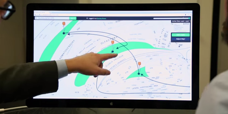
A study conducted by a London based behavioural research consultancy has collected some interesting data about the brain activity of poker players. The study set out to record the levels of brain activity in poker players operating at different levels of competence: the beginner, the amateur and the professional. Six players, two from each category, were observed playing forty minutes of Texas Hold’em poker. Half played for money, half for free. The players wore EEG headsets which recorded the location and intensity of brain activity in four areas of the brain: delta, theta, alpha and beta. This data was then converted into interactive brain maps, allowing us to observe brain activity in the players at key moments of the game.

Deal


Brain Maps of Poker Players

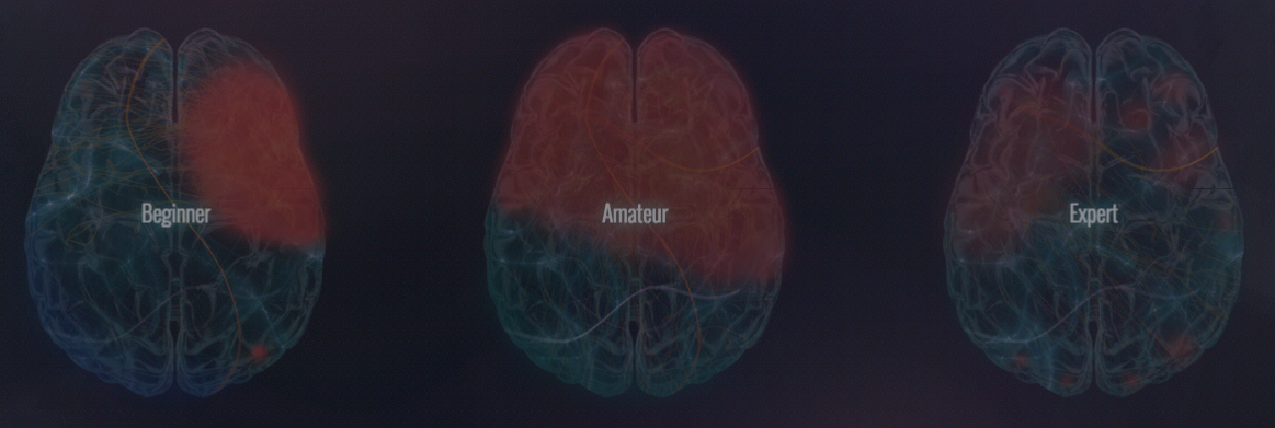
At the start of the game the beginner shows high theta activity in the right frontal lobe, indicating high levels of emotion, in contrast, the amateur displays high beta activity in the left frontal lobe, indicating decision making driven by logic. This is the stage of the game where the amateur is most engaged and where they spend the most time processing the information. The brain map of the professional is similar but indicates a much lower level of activity. The experienced player makes a quicker decision with less mental effort.


Flop


Related articles
This is where the first three cards are placed down altogether, face up. The beginner’s brain map shows little activity. Their lack of experience renders them incapable of responding to the new data, either with emotion or logic. The amateur shows alpha activity indicating logic at work but both brain maps are in stark contrast to that of the professional where a high level of activity in both frontal lobes indicates both logical thinking and emotional instinct at work.

River


This is a key phase of the game when the fifth card is placed, face up. The beginner’s brain exhibits exclusive right frontal lobe activity, indicating an entirely emotional response to the situation. The amateur brain map shows high levels of activity in both frontal lobes, with slightly more activity on the right lobe, suggesting that emotion is dominant. The effect of the final card on the professional is to stimulate a flurry of activity on the left lobe. The professional is in control of emotion and is relying on logic to make the decision.

Call


This is when a player adds to the pot, money equal to the most recent bet. The fact that this is a relatively safe play is reflected in similar brain maps for all three subjects. As we might expect, the response of the beginner is predominantly emotional whereas the brain maps for the amateur and professional show more of a spread across both frontal lobes.

Raise

Excitement peaks for all players when the stakes are raised and this is evidenced by the brain maps. What is also revealed is that the professional, although led by emotion, has far more brain activity devoted to processing information.


By  33rd SquareEmbed


Sunday, September 18, 2016

Human Brain Atlas Updated with Unprecedented Resolution


Neuroscience

The new Allen Brain Atlas combines neuroimaging and tissue staining to offer an unprecedented level of resolution. The high-resolution brain map features 862 brain structures from a single donor brain.


Researchers at the Allen Brain Institute, have developed the first whole human brain map at cellular resolution. The dataset combines neuroimaging and comprehensive mapping of brain regions. This digital human brain atlas will allows researchers to investigate the structural basis of human brain function at a resolution never possible before.

The Allen Human Brain Atlas is a unique multi-modal atlas that maps gene expression across the human brain. The model integrates anatomic and genomic information, available data modalities include magnetic resonance imaging (MRI), diffusion tensor imaging (DTI), histology, and gene expression data derived from both microarray and in situ hybridization (ISH) approaches.


"Essentially what we were trying to do is to create a new reference standard for a very fine anatomical structural map of the complete human brain."
Some of the key features include an "all genes, all structures" microarray survey spatially mapped to the MRI, ISH cellular resolution image data for selected genes in specific brain regions, and an annotated human brain atlas guide.

Now with the latest updates to the Human Brain Atlas, users can navigate the brain as easily as using Google Maps. The work was published recently in The Journal of Comparative Neurology.

Related articles
“Essentially what we were trying to do is to create a new reference standard for a very fine anatomical structural map of the complete human brain,” says Ed Lein, principal investigator on the project. “It may seem a little bit odd, but actually we are a bit lacking in types of basic reference materials for mapping the human brain that we have in other organisms like mouse or like monkey, and that is in large part because of the enormous size and complexity of the human brain.”

The project, which has already been running for five years, focused on a single healthy postmortem brain from a 34-year-old woman. The research team took a complete scan of the organ, which allowed them to capture both overall brain structure and the connectivity of brain fibers.

The researchers next sliced the brain into 2,716 very thin sections for fine-scale, cellular analysis.

The key step in creating a complete brain atlas was combining broad-scale, high-resolution brain imaging data with detailed cellular-level mapping, which the researchers then annotated with the brain structures they identified. This work is now publically available as the Allen Human Brain Atlas.

"Because of the labor intensiveness of doing this, it always lives in the scale of a single brain,” Lein says, “and you really go to town in trying to understand everything you can about that one individual."

Other researchers argue that the work is too discrete, being from one subject, and that it will be inconclusive to make any broad ranging neuroscience observations from the data.

A different map of the brain was published earlier this year by the Human Connectome Project. The main difference is the Allen study used only one brain, while the Human Connectome Project mapped 210 brains.

In spite of the critics,  the effort marks a marked advance in our understanding of brain anatomy. “There simply hasn't been a complete map of the human brain as a reference piece of material for anyone studying any part of the brain,” Lein says, “and this is a completely essential part of doing research.”

The Allen Human Brain Atlas features anatomic and gene-based search options as well as interactive viewing with the Brain Explorer 3D software.


SOURCE  Scientific American, Allen Brain Institute


By  33rd SquareEmbed


Wednesday, October 22, 2014


 Neuroscience
Neuroscience researchers have created Golgi, an interactive map of a rat brain that makes exploring the brain as easy as using Google Maps.


What is the link in your brain between smell and memory? Where’s the connection between habits and Parkinson’s disease? How does one detour into addiction?

To answer these and other complex scientific and medical questions, two University of Southern California scientists have built Golgi, an interactive map of a rat brain that makes exploring the brain as easy as using Google Maps.

The map can be found online at www.useGolgi.com.

www.useGolgi.com


"We have a big advantage because we’re the only group — really in the world — that has a flat map of the brain."


The new Web app, unveiled today, offers details at the click of a button about how the regions of the brain communicate and interact. Golgi will help accelerate the research and treatment of diseases like Parkinson’s and depression by layering complex scientific data onto a single simple brain map that provides information to doctors and researchers quickly and intuitively.

“We have a big advantage because we’re the only group — really in the world — that has a flat map of the brain,” said Larry Swanson, National Academy member, professor of biological sciences at the USC Dornsife College of Letters, Arts and Sciences and recent past president of the Society for Neuroscience.

Swanson, a longtime pillar of the neuroscience community, collaborated with 25-year-old USC graduate student Ramsay Brown, who designed the program as an undergraduate worker in Swanson’s lab.

Related articles
Golgi takes the existing pool of knowledge about the brain and makes it easy to access by scientists and doctors, saving time and offering potentially new connections. It uses research on rat brains, which are close enough to human brains to offer valuable insights but are far easier to study and therefore represent a larger pool of research data.

To display the brain’s three-dimensional structure on two-dimensional screens, Swanson and Brown used the embryonic brain — which begins as a flat sheet of cells — as a guide. This flattens the brain and keeps related portions of the brain located close together. Flattening the brain lets users click around and display connectome and other data directly on regions they’re interested in learning about for research or treatment.

“We designed a really intuitive way to explore the more nuanced details about the brain and connectome,” Brown said. “Making this data easy and accessible will improve how scientists and doctors explore, explain and treat human conditions and restore quality of life — and that’s really special to us.”

Brown and Swanson think this program is just the beginning.

Connectomics, the subfield of neuroscience that studies and maps the brain’s wiring, is advancing quickly and providing better maps as the technique evolves. Programs like Golgi will help doctors and researchers make sense of these new maps and make better medical and scientific decisions faster.

“Many people now think that understanding these neurological diseases is going to require understanding the circuitry of the brain,” Swanson said.


SOURCE  USC

By 33rd SquareEmbed

Tuesday, October 21, 2014

Unlocking the Power of the Mind - Up and Coming Medical Tech

 Medicine
The human brain remains the most complex phenomenon in the known universe. More and more, medical research is uncovering the mysteries of the brain and applying that knowledge to new technology.


In the last few years, medical practice and the growing complexity and refinement of technology has led to some astounding breakthroughs in a number of medical fields. Interestingly, while technology has certainly been responsible for some groundbreaking implementations on its own, it is often the ability to understand the inner workings of the human body through technology that allow medical science to make huge leaps forward in preventative and curative medical approaches. The human brain is often at the forefront of such inquiry, as in many ways remains the last true enigma.

Man-Machine Interface

Some of the most impressive breakthroughs in recent times have undoubtedly been the ability to harness brain waves, in turn using them to enable control of tech. Perhaps one of the most arresting events is the recent story of a Swedish truck driver, who lost an arm ten years ago. Through a process they named ‘Osseointergration’, scientists at Chalmers University of Technology created an interface between the biological and technological allow the man to have a much wider level of control over his new prosthetic arm. While this kind of basic idea has already been achieved by placing electrodes on patients, this direct link between man and machine is the first of its kind, and opens up a huge amount of possibilities for the future.

Related articles
Work has already begun on finding ways to allow signals to travel back from the prosthetic to the brain, in an effort to enable to the user to essentially ‘feel’ their arm. This goes hand in hand with recent breakthroughs in the US, which allow prosthetic arms to be controlled through the electrical signals of muscle contractions. Of course, behind these amazing innovations is the application of computer models and software - in essence technology and medicine are now working more closely than ever before. As we gain an ever increasing understanding of the brain, the way we approach medical problems will begin to, and already has in some cases, begin to change forever.

Understanding how we process information for example, has led to understanding how to implement accelerated learning, and the ongoing research into addiction disorders is also looking promising. Even simple smartphone apps are having a positive impact, especially when used in addition to traditional therapy and support.

Mapping the Brain

Ongoing research of the brain has led to a number of other recent discoveries, which set the stage for some truly exciting applications in the near future. Brain Mapping itself has been ongoing for a decade, and as a result scientists and neuroscientists are now able to view the brain in higher detail than ever before.

brain mapping

Perhaps what is even more exciting however, is how the relationship between the organic and the technological continue to evolve in a way that mirror each other. Neuromorphic chips for example, are modeled on the brain itself, and have to potential to give computers much more awareness and power - something that will be fundamental in the ongoing development of robots and artificial intelligence. The chips are not set to be manufactured until next year, but the prospects are certainly exciting. The basic idea behind the chip is that it uses silicon to model the neural pathways of the brain. This in turn allows the chip to not only achieve much higher processing speeds, but also helps to circumvent the traditional problems that processors currently face: heat.

Despite continual advances in tech, a processor as we know it today are consistently limited by heat generation. The neuromorphic chip could bypass this problem, potentially leading to some major new advancements in both computer power and speed, but also in the range and ability of tasks a computer will be able to handle.

While it may be some time before the Qualcomm developed chips are sitting comfortably in our smartphones and tablets, the potential benefits for almost all areas of the computer tech industry and others is huge. In the end, despite our increasingly and ever growing technical savvy, the neuromorphic chip serves as a great example of how we can unlock better, more efficient tech by unlocking more about ourselves and the natural world around us.


SOURCE  Top Image - Human Connectome Project

By Anne FoyEmbed

Thursday, October 2, 2014

High Resolution Electrode Array System Will Help Us Understand The Brain

 Neuroscience
As part of the BRAIN Initiative, researchers are developing an electrode array system that will “enable researchers to better understand how the brain works through unprecedented resolution and scale.”


The American National Institutes of Health (NIH) awarded Lawrence Livermore National Laboratory (LLNL) a grant to develop an electrode array system that will enable researchers to better understand how the brain works through unprecedented resolution and scale.

LLNL's grant-funded project is part of NIH's efforts to support President Obama's BRAIN (Brain Research through Advancing Innovative Neurotechnologies) Initiative, a new research effort to revolutionize our understanding of the human mind and uncover ways to treat, prevent and cure brain disorders. NIH is seeking exceptionally creative approaches to address major challenges associated with recording and manipulating neural activity for this endeavor.

"There's a big gap between what we want to do in brain research and the technologies available to make exploration possible. These initial awards are part of a 12-year scientific plan focused on developing the tools and technologies needed to make the next leap in understanding the brain. This is just the beginning of an ambitious journey and we're excited about the possibilities."


The agency announced its first wave of investments totaling $46 million in FY14 funds to support the BRAIN Initiative's goals. More than 100 investigators in 15 states and several countries will work to develop new tools and technologies to understand neural circuit function and capture a dynamic view of the brain in action.

"The human brain is the most complicated biological structure in the known universe. We've only just scratched the surface in understanding how it works -- or, unfortunately, doesn't quite work when disorders and disease occur," NIH Director Francis S. Collins said. "There's a big gap between what we want to do in brain research and the technologies available to make exploration possible. These initial awards are part of a 12-year scientific plan focused on developing the tools and technologies needed to make the next leap in understanding the brain. This is just the beginning of an ambitious journey and we're excited about the possibilities."


neural measurement and manipulation system

Lawrence Livermore is developing a neural measurement and manipulation system -- an advanced electronics system to monitor and modulate neurons -- that will be packed with more than 1,000 tiny electrodes embedded in different areas of the brain to record and stimulate neural circuitry. The goal is to develop a system that will allow scientists to simultaneously study how thousands of neuronal cells in various brain regions work together during complex tasks such as decision making and learning.

The biologically compatible neural system will be the first of its kind to have large-scale network recording capabilities that are designed to continuously record neural activities for months to years.

"This is an incredible opportunity for us to develop a technology that is going to advance neuroscience research for the community," said Vanessa Tolosa, an engineer at LLNL's Center for Bioengineering who is a principal investigator on the project. "The brain is a dynamic and complicated system. Though neuroscientists have uncovered a lot about the brain in the last couple of decades, there is a pressing need for new technologies that'll enable us to study more brain regions over longer periods of time."

The NIH project is a collaboration between LLNL's Neural Technology Group; the laboratory of Loren Frank at University of California, San Francisco (UCSF); Intan Technology; and SpikeGadgets.

Housed at the Center for Bioengineering, the Neural Technology Group will work with UCSF researchers to design and build electrode arrays that can record hundreds to thousands of brain cells simultaneously. Their goal is to develop 1,000-plus channel arrays that can eventually be expanded to 10,000 channels.

Related articles
These arrays will use new microchips designed at Intan and will send data to a system developed at SpikeGadgets. UCSF will coordinate these efforts and test the technologies. The arrays will penetrate multiple regions of the brain without interfering with normal functions during the experiments, allowing for detailed studies of brain circuits that underlie behavior.

"This collaboration combines the engineering talent of LLNL with UCSF's expertise in neural recording and modulation systems, and the design and programming skills of Intan and SpikeGadgets," Frank said. "The result will be a system that will help us understand how different brain areas communicate and carry out complex mental functions."

The system also will be designed for compatibility with optogenetic stimulation, a technique that uses light sensitive proteins and light to manipulate neural activity. This technique allows researchers to target specific neurons or cells for recording.

Using the Center for Bioengineering's unique microfabrication capabilities, Tolosa and her colleagues have achieved multiple patents and publications during the last decade. The team's ultimate goal is to launch a complete, modular and open source system that would be available to any neuroscientists interested in large-scale neural recording and modulation.

LLNL's project is one of 58 BRAIN-related projects funded by NIH. The others include creating a wearable scanner to image the human brain in motion, using lasers to guide nerve cell firing, recording the entire nervous system in action, stimulating specific circuits with radio waves and identifying complex circuits with DNA barcodes. The majority of the NIH grants focus on developing transformative technologies that will accelerate fundamental neuroscience research and include:
  • -Classifying the myriad cell types in the brain
  • -Producing tools and techniques for analyzing brain cells and circuits
  • -Creating next-generation human brain imaging technology
  • -Developing methods for large-scale recordings of brain activity
  • -Integrating experiments with theories and models to understand the functions of specific brain circuits

SOURCE  Lawrence Livermore National Laboratory

By 33rd SquareEmbed

Monday, September 8, 2014

Researchers Looking To Map The Brain Find It Even More Complex Than They Thought

 Neuroscience
Researchers have found that the brain's wiring is more complex than expected – one set of neural wires can trigger different reactions, depending on how it fires. The work opens new questions for scientists trying to map the brain's connections.


Scientists at Stanford's Bio-X have raised an entirely new set of questions when they sought answers about connections between two brain regions.

"There's a lot of excitement about being able to make a map of the brain with the idea that if we could figure out how it is all connected we could understand how it works,"  researcher Joanna Mattis said. "It turns out it's so much more dynamic than that."

Mattis is a co-first author on a paper describing the work published recently in the Journal of Neuroscience. Julia Brill, then a postdoctoral scholar, was the other co-first author.

Mattis had been a graduate student in the lab of Karl Deisseroth, professor of bioengineering and of psychiatry and behavioral sciences, where she helped work on a new technique called optogenetics. That technique allows neuroscientists to selectively turn parts of the brain on and off to see what happens. She wanted to use optogenetics to understand the wiring of a part of the brain involved in spatial memory – it's what makes a mental map of your surroundings as you explore a new city, for example.

Related articles
Scientists already knew that when an animal explores habitats, two parts of the brain are involved in the initial exploring phase and then in solidifying a map of the environment – the hippocampus and the septum.

When an animal is exploring an environment, the neurons in the hippocampus fire slow signals to the septum, essentially telling the septum that it's busy acquiring information. Once the animal is done exploring, those same cells fire off intense signals letting the septum know that it's now locking that information into memory. The scientists call this phase consolidation. The septum uses that information to then turn around and regulate other signals going into the hippocampus.

"I wanted to study the hippocampus because on the one hand so much was already known – there was already this baseline of knowledge to work off of. But then the question of how the hippocampus and septum communicate hadn't been accessible before optogenetics," Mattis said.

connectome
Neural wiring diagrams are even more complex than initially speculated according to new research
Neurons in the hippocampus were known to fire in a rhythmic pattern, which is a particular expertise of John Huguenard, a professor of neurology. Mattis obtained an interdisciplinary fellowship through Stanford Bio-X, which allowed her to combine the Deisseroth lab's expertise in optogenetics with the rhythmic brain network expertise of Julia Brill from the Huguenard lab.

"There's a lot of excitement about being able to make a map of the brain with the idea that if we could figure out how it is all connected we could understand how it works."


Mattis and Brill used optogenetics to prompt neurons of the hippocampus to mimic either the slow firing characteristic of information acquisition or the rapid firing characteristic of consolidation. When they mimicked the slow firing they saw a quick reaction by cells in the septum. When they mimicked the fast consolidation firing, they saw a much slower response by completely different cells in the septum.

Same set of wires – different outcome. That's like turning on different lights depending on how hard you flip the switch. "This illustrates how complex the brain is," Mattis said.

This research has raised a whole new set of questions, Mattis said. They more or less understand the faster reaction, but what is causing the slower reaction? How widespread is this phenomenon in the brain?

"The other big picture thing that we opened up but didn't answer is: How can you then tie this back to the circuit overall and learning memory?" Mattis said. "Those would be exciting things to follow up on for future projects."


SOURCE  Stanford

By 33rd SquareEmbed

Monday, June 9, 2014

cholinergic neuron mapping

 Neuroscience
By studying laboratory mice, scientists at The Johns Hopkins University have succeeded in plotting the labyrinthine paths of some of the largest nerve cells in the mammalian brain: cholinergic neurons, the first cells to degenerate in people with Alzheimer's disease.  


Scientists at The Johns Hopkins University have succeeded in plotting the labyrinthine paths of some of the largest nerve cells in the mammalian brain: cholinergic neurons, the first cells to degenerate in people with Alzheimer’s disease.

“For us, this was like scaling Mount Everest,” says Jeremy Nathans, Ph.D., professor of molecular biology and genetics, neuroscience, and ophthalmology at the Johns Hopkins University School of Medicine.

"We think something like this might be happening when cholinergic neurons trying to repair the damage done by Alzheimer’s disease."


“This work reveals the amazing challenges that cholinergic neurons face every day. Each of these cells is like a city connected to its suburbs by a single, one-lane road, with all of the emergency services located only in the city. You can imagine how hard it would be in a crisis if all of the emergency vehicles had to get to the suburbs along that one road. We think something like this might be happening when cholinergic neurons trying to repair the damage done by Alzheimer’s disease.”

Each cholinergic neuron, Nathans explains, has roughly 1,000 branch points. If lined up end to end, one neuron’s branches would add up to approximately 15 times the length of the mouse brain. But all of the branches are connected by a single, extremely thin “pipeline” to one hub — the cell body — that provides for the needs of the branches. The challenge of moving material through this single pipeline could make it very difficult for cholinergic neurons to combat the challenges that come with a disorder like Alzheimer’s disease, he says. Now, by mapping the branches and pipelines, scientists will likely get a better fix on what happens when the neurons fail to meet the challenges.

A summary of the research was published online in the journal eLife.

Cholinergic Neurons

Related articles
Cholinergic neurons are among the largest neurons in the mammal brain. Named for their release of a chemical messenger called acetylcholine, they number only in the thousands in mouse brains, a tiny fraction of the 50 to 100 million total neurons. Their cell bodies are located at the base of the brain near its front end, but their branches extend throughout the cerebral cortex, the outermost, wrinkled layer of “grey matter” that is responsible for the mind’s most advanced intellectual functions. Therefore, although there are relatively few cholinergic neurons, they affect a very large part of the brain, Nathans says.

Due to the technical challenge of visualizing the complicated paths of hundreds of tiny branches from a single neuron tangled within millions of other neurons, the actual size and shape of individual cholinergic neurons — and the territory they cover — had been unknown until now, Nathans says. Using genetic engineering methods, the Nathans team programmed several cholinergic neurons per mouse to make a protein that could be seen with a colored chemical reaction.

Critical to the success of the work was the ability to limit the number of cells making the protein — if all of the cholinergic neurons made the protein, it would have been impossible to distinguish individual branches.

Because microscopes cannot see through thick tissue, Nathans and his team preserved the mouse brains and then thinly sliced them to produce serial images. The branching path of each neuron was then painstakingly reconstructed from the serial images and analyzed. In adult mice, he says, the average length of the branches of a single cholinergic neuron, lined up end to end, is 31 cm (12 inches), varying from 11 to 49 cm (4 to 19 inches). The average length of a mouse brain is only 2 cm — a bit less than one inch. Although each cholinergic neuron, on average, contains approximately 1,000 branch points, they vary significantly in the extent of the territory that they cover.

The researchers used the same techniques to study the cholinergic neurons of mice with a rodent form of Alzheimer’s disease and found that the branches were fragmented. They also found clumps of material that may have been debris from the disintegrating branches.

Although the cholinergic neurons of human brains have not been individually traced, Nathans’ team was able to calculate that the average cholinergic neuron in the human brain has a total branch length of approximately 100 meters, a bit longer than a football field. “That is a really long pipeline, especially if one considers that the pipes have diameters of only 30 thousandths of a millimeter, far narrower than a human hair,” says Nathans.

He adds, “Although our study only defined a few simple, physical properties of these neurons, such as size and shape, it has equipped us to form and test better hypotheses about what goes wrong with them during disease.”


SOURCE  Johns Hopkins University School of Medicine

By 33rd SquareEmbed

Thursday, April 3, 2014

Mouse Connectome

 Neuroscience
Scientists from the Allen Institute for Brain Science in Seattle have built two new brain maps: one of gene expression in the developing human brain, and another of neural networks in a mouse brain. The maps, which are publicly available, will serve as resources for researchers around the world.


Researchers from the Allen Institute for Brain Science have published the first comprehensive, large-scale data set on how the brain of a mammal is wired, providing a groundbreaking data resource and fresh insights into how the nervous system processes information. Their landmark paper in the journal Nature both describes the publicly available Allen Mouse Brain Connectivity Atlas, and demonstrates the exciting knowledge that can be gleaned from this valuable resource.

“Understanding how the brain is wired is among the most crucial steps to understanding how the brain encodes information,” explains Hongkui Zeng, Senior Director of Research Science at the Allen Institute for Brain Science. “The Allen Mouse Brain Connectivity Atlas is a standardized, quantitative, and comprehensive resource that will stimulate exciting investigations around the entire neuroscience community, and from which we have already gleaned unprecedented details into how structures are connected inside the brain.”

Using the data, Allen Institute scientists were able to demonstrate that there are highly specific patterns in the connections among different brain regions, and that the strengths of these connections vary with greater than five orders of magnitudes, balancing a small number of strong connections with a large number of weak connections. This publication comes just as the research team wraps up more than four years of work to collect and make publicly available the data behind the Allen Mouse Brain Connectivity Atlas project, with the completion of the Atlas announced in March 2014.

Mouse Brain Atlas

"The kind of analysis we have done so far is just the beginning of the deep analysis of the wiring patterns of different brain circuits made possible by this unique collection of data."


The human brain is among the most complex structures in the entire universe, containing roughly 100 billion neurons—as many stars as are in the Milky Way. The mouse brain’s 75 million neurons, arranged in a roughly similar structure to the human brain, provide a powerful model system by which to understand how nerve cells of the human brain connect, process and encode information.

Despite the foundational need to understand how areas of the brain are connected, the only species for which we have a complete wiring diagram is the simple microscopic worm C. elegans—a far simpler system, with only 302 neurons, compared to the human or any other mammalian nervous system.

Scientists at the Allen Institute set out to create a wiring diagram of the brain—also known as a connectome—to illustrate short and long-range connections using genetically-engineered viruses that could trace and illuminate individual neurons. In order to get a truly comprehensive view, scientists collected imaging data at resolutions smaller than a micrometer from more than 1,700 mouse brains, each of which was divided into 140 serial sections. “The data for the Allen Mouse Brain Connectivity Atlas was collected in a way that’s never been done before,” says Zeng. “Standardizing the data generation process allowed us to create a 3D common reference space, meaning we could put the data from all of our thousands of experiments next to each other and compare them all in a highly quantitative way at the same time.”

The Allen Mouse Brain Connectivity Atlas contains more than 1.8 petabytes of data—the equivalent of 23.9 years of continuous HD video—all of which is freely available online to the entire community. The research team behind the Atlas has been steadily releasing new data since November 2011; and in March, they released the last major update to the Atlas, though the resource will continue to be updated as technology develops and researchers are able to add more new types of connectivity data. Like all of the Allen Brain Atlas resources, the data and the tools to browse and analyze them are freely available to the public at www.brain-map.org.

As a freely available resource, the Allen Mouse Brain Connectivity Atlas is an invaluable tool for neuroscientists with questions about the nature of the brain’s connections.

"The Allen Mouse Brain Connectivity Atlas provides an initial road-map of the brain, at the level of interstate highways and the major cities that they link,” explains David Anderson, Professor of Biology and Howard Hughes Medical Institute Investigator at the California Institute of Technology. “Smaller road networks and their intersections with the interstates will be the next step, followed by maps of local streets in different municipalities. This information will provide a framework for what we ultimately want to understand: ‘traffic patterns’ of information flow in the brain during various activities such as decision-making, mapping of the physical environment, learning and remembering, and other cognitive or emotional processes."

Related articles
With the Nature publication, Allen Institute scientists have already begun to demonstrate the power of analysis contained within the Atlas. By analyzing the data, Zeng and her team were able to discover several interesting properties of the mouse brain’s connectome. For example, there are extensive connections across the two hemispheres with mirror-image symmetry. Pathways belonging to different functional circuits in the brain can be identified and their relationships and intersections visualized in 3D. Finally, there is a great degree of variation in the strengths of all the connections–ranging beyond five orders of magnitude—and an intriguing balance between a small number of strong connections and a large number of weak connections.

These discoveries illustrate the need for a quantitative understanding and a global view of the brain’s connectivity patterns, since a quantitative approach can describe the relative strength of different connections instead of the simple presence or absence descriptions that are inherent to a more qualitative approach. These more accurate comparisons are uniquely enabled by the Atlas, Zeng says.

“The purpose of the Atlas is to create a new way to map the brain’s vast connections systematically and rapidly, and to develop a platform to present the data to users and help them navigate in the friendliest possible way,” explains Zeng. “But the kind of analysis we have done so far is just the beginning of the deep analysis of the wiring patterns of different brain circuits made possible by this unique collection of data.”

Maintaining the Allen Mouse Brain Connectivity Atlas is a continuous effort. After the completion of the Atlas as originally scoped in March 2014, scientists will continue to update the Atlas with profiles of more individual nerve cell types as they become available. Researchers at the Allen Institute are also poised to dive more deeply into the data they have already collected, and will focus more intently on studying the connections between different types of neurons in the same or neighboring regions – the city roads and local streets that, together with the interstates, form the hierarchical neural networks.

“Who you are—all your thoughts and actions your entire life—is based on connections between neurons,” explains Ed Callaway, Professor in the Systems Neurobiology Laboratories at the Salk Institute for Biological Studies. “So if we want to understand any of these processes or how they go wrong in disease, we have to understand how those circuits function. Without an atlas, we couldn’t hope to gain that understanding.”


SOURCE  Allen Institute for Brain Science

By 33rd SquareEmbed

Wednesday, February 12, 2014

Research Uncovers White Matter Scaffold of Human Brain

 Neuroscience
For the first time, neuroscientists have systematically mapped the white matter "scaffold" of the human brain, the critical communications network that supports brain function.


For the first time, neuroscientists have systematically identified the white matter "scaffold" of the human brain, the critical communications network that supports brain function.

Their work, published in the open-source journal Frontiers in Human Neuroscience, has major implications for understanding brain injury and disease. By detailing the connections that have the greatest influence over all other connections, the researchers offer not only a landmark first map of core white matter pathways, but also show which connections may be most vulnerable to damage.

"We coined the term white matter 'scaffold' because this network defines the information architecture which supports brain function," said senior author John Darrell Van Horn of the USC Institute for Neuroimaging and Informatics and the Laboratory of Neuro Imaging at USC.

"While all connections in the brain have their importance, there are particular links which are the major players," Van Horn said.

Graphical representation of human brain connectivity scaffold
Graphical representation of human brain connectivity scaffold.
Image Source -  USC Institute for Neuroimaging and Informatics
Using MRI data from a large sample of 110 individuals, lead author Andrei Irimia, also of the USC Institute for Neuroimaging and Informatics, and Van Horn systematically simulated the effects of damaging each white matter pathway.

Related articles
They found that the most important areas of white and gray matter don't always overlap. Gray matter is the outermost portion of the brain containing the neurons where information is processed and stored. Past research has identified the areas of gray matter that are disproportionately affected by injury.

But the current study shows that the most vulnerable white matter pathways – the core "scaffolding" – are not necessarily just the connections among the most vulnerable areas of gray matter, helping explain why seemingly small brain injuries may have such devastating effects.

"Sometimes people experience a head injury which seems severe but from which they are able to recover. On the other hand, some people have a seemingly small injury which has very serious clinical effects," says Van Horn, associate professor of neurology at the Keck School of Medicine of USC. "This research helps us to better address clinical challenges such as traumatic brain injury and to determine what makes certain white matter pathways particularly vulnerable and important."

The researchers compare their brain imaging analysis to models used for understanding social networks. To get a sense of how the brain works, Irimia and Van Horn did not focus only on the most prominent gray matter nodes – which are akin to the individuals within a social network. Nor did they merely look at how connected those nodes are.

white matter brain connections
Image Source -  USC Institute for Neuroimaging and Informatics
Rather, they also examined the strength of these white matter connections, i.e. which connections seemed to be particularly sensitive or to cause the greatest repercussions across the network when removed. Those connections which created the greatest changes form the network "scaffold."

"Just as when you remove the internet connection to your computer you won't get your email anymore, there are white matter pathways which result in large scale communication failures in the brain when damaged," Van Horn said.

When white matter pathways are damaged, brain areas served by those connections may wither or have their functions taken over by other brain regions, the researchers explain. Irimia and Van Horn's research on core white matter connections is part of a worldwide scientific effort to map the 100 billion neurons and 1,000 trillion connections in the living human brain, led by the Human Connectome Project and the Laboratory of Neuro Imaging at USC.

Irimia notes that, "these new findings on the brain's network scaffold help inform clinicians about the neurological impacts of brain diseases such as multiple sclerosis, Alzheimer's disease, as well as major brain injury. Sports organizations, the military and the US government have considerable interest in understanding brain disorders, and our work contributes to that of other scientists in this exciting era for brain research."


SOURCE  University of Southern California

By 33rd SquareSubscribe to 33rd Square

Tuesday, October 15, 2013

Mind-Reading Devices May Be On the Horizon According to Stanford Scientists

 Mind Reading
Researchers have found the first solid evidence that the pattern of brain activity seen in someone performing a mathematical exercise under experimentally controlled conditions is very similar to that observed when the person engages in thinking about numbers and quantities in general.


A brain region activated when people are asked to perform mathematical calculations in an experimental setting is similarly activated when they use numbers — or even imprecise quantitative terms, such as “more than”— in everyday conversation, according to a study by Stanford University School of Medicine scientists.

Using a new method, the researchers collected the first solid evidence that the pattern of brain activity seen in someone performing a mathematical exercise under experimentally controlled conditions is very similar to that observed when the person engages in quantitative thought in the course of daily life.

“We’re now able to eavesdrop on the brain in real life,” said Josef Parvizi, MD, PhD, associate professor of neurology and neurological sciences and director of Stanford’s Human Intracranial Cognitive Electrophysiology Program. Parvizi is the senior author of the study, published in Nature Communications. The study’s lead authors are postdoctoral scholar Mohammad Dastjerdi, MD, PhD, and graduate student Muge Ozker.

The finding could lead to “mind-reading” applications that, for example, would allow a patient who is rendered mute by a stroke to communicate via passive thinking. Conceivably, it could also lead to more dystopian outcomes: chip implants that spy on or even control people’s thoughts.

“This is exciting, and a little scary,” said Henry Greely, JD, the Deane F. and Kate Edelman Johnson Professor of Law and steering committee chair of the Stanford Center for Biomedical Ethics, who played no role in the study but is familiar with its contents and described himself as “very impressed” by the findings. “It demonstrates, first, that we can see when someone’s dealing with numbers and, second, that we may conceivably someday be able to manipulate the brain to affect how someone deals with numbers.”

The researchers monitored electrical activity in a region of the brain called the intraparietal sulcus, known to be important in attention and eye and hand motion. Previous studies have hinted that some nerve-cell clusters in this area are also involved in numerosity, the mathematical equivalent of literacy.

However, the techniques that previous studies have used, such as functional magnetic resonance imaging, are limited in their ability to study brain activity in real-life settings and to pinpoint the precise timing of nerve cells’ firing patterns. These studies have focused on testing just one specific function in one specific brain region, and have tried to eliminate or otherwise account for every possible confounding factor. In addition, the experimental subjects would have to lie more or less motionless inside a dark, tubular chamber whose silence would be punctuated by constant, loud, mechanical, banging noises while images flashed on a computer screen.

Related articles
“This is not real life,” said Parvizi. “You’re not in your room, having a cup of tea and experiencing life’s events spontaneously.” A profoundly important question, he said, is: “How does a population of nerve cells that has been shown experimentally to be important in a particular function work in real life?”

His team’s method, called intracranial recording, provided exquisite anatomical and temporal precision and allowed the scientists to monitor brain activity when people were immersed in real-life situations. Parvizi and his associates tapped into the brains of three volunteers who were being evaluated for possible surgical treatment of their recurring, drug-resistant epileptic seizures.

The procedure involves temporarily removing a portion of a patient’s skull and positioning packets of electrodes against the exposed brain surface. For up to a week, patients remain hooked up to the monitoring apparatus while the electrodes pick up electrical activity within the brain. This monitoring continues uninterrupted for patients’ entire hospital stay, capturing their inevitable repeated seizures and enabling neurologists to determine the exact spot in each patient’s brain where the seizures are originating.

During this whole time, patients remain tethered to the monitoring apparatus and mostly confined to their beds. But otherwise, except for the typical intrusions of a hospital setting, they are comfortable, free of pain and free to eat, drink, think, talk to friends and family in person or on the phone, or watch videos.

The electrodes implanted in patients’ heads are like wiretaps, each eavesdropping on a population of several hundred thousand nerve cells and reporting back to a computer.

In the study, participants’ actions were also monitored by video cameras throughout their stay. This allowed the researchers later to correlate patients’ voluntary activities in a real-life setting with nerve-cell behavior in the monitored brain region.

As part of the study, volunteers answered true/false questions that popped up on a laptop screen, one after another. Some questions required calculation — for instance, is it true or false that 2+4=5? — while others demanded what scientists call episodic memory — true or false: I had coffee at breakfast this morning. In other instances, patients were simply asked to stare at the crosshairs at the center of an otherwise blank screen to capture the brain’s so-called “resting state.”

Consistent with other studies, Parvizi’s team found that electrical activity in a particular group of nerve cells in the intraparietal sulcus spiked when, and only when, volunteers were performing calculations.

Afterward, Parvizi and his colleagues analyzed each volunteer’s daily electrode record, identified many spikes in intraparietal-sulcus activity that occurred outside experimental settings, and turned to the recorded video footage to see exactly what the volunteer had been doing when such spikes occurred.

They found that when a patient mentioned a number — or even a quantitative reference, such as “some more,” “many” or “bigger than the other one” — there was a spike of electrical activity in the same nerve-cell population of the intraparietal sulcus that was activated when the patient was doing calculations under experimental conditions.

That was an unexpected finding. “We found that this region is activated not only when reading numbers or thinking about them, but also when patients were referring more obliquely to quantities,” said Parvizi.

“These nerve cells are not firing chaotically,” he said. “They’re very specialized, active only when the subject starts thinking about numbers. When the subject is reminiscing, laughing or talking, they’re not activated.” Thus, it was possible to know, simply by consulting the electronic record of participants’ brain activity, whether they were engaged in quantitative thought during nonexperimental conditions.

Any fears of impending mind control are, at a minimum, premature, said Greely. “Practically speaking, it’s not the simplest thing in the world to go around implanting electrodes in people’s brains. It will not be done tomorrow, or easily, or surreptitiously.”

Parvizi agreed. “We’re still in early days with this,” he said. “If this is a baseball game, we’re not even in the first inning. We just got a ticket to enter the stadium.”